Dissection of jejunum, ileum and colon
Lymphatic structures, blood vessels and nerves of lower part of sigmoid colon, close-up view
Stanford holds the copyright to the David L. Bassett anatomical images and has assigned
Creative Commons license Attribution-Share Alike 4.0 International to all of the images.
For additional information regarding use and permissions,
please contact Dr. Drew Bourn at dbourn@stanford.edu.
Image #144-4
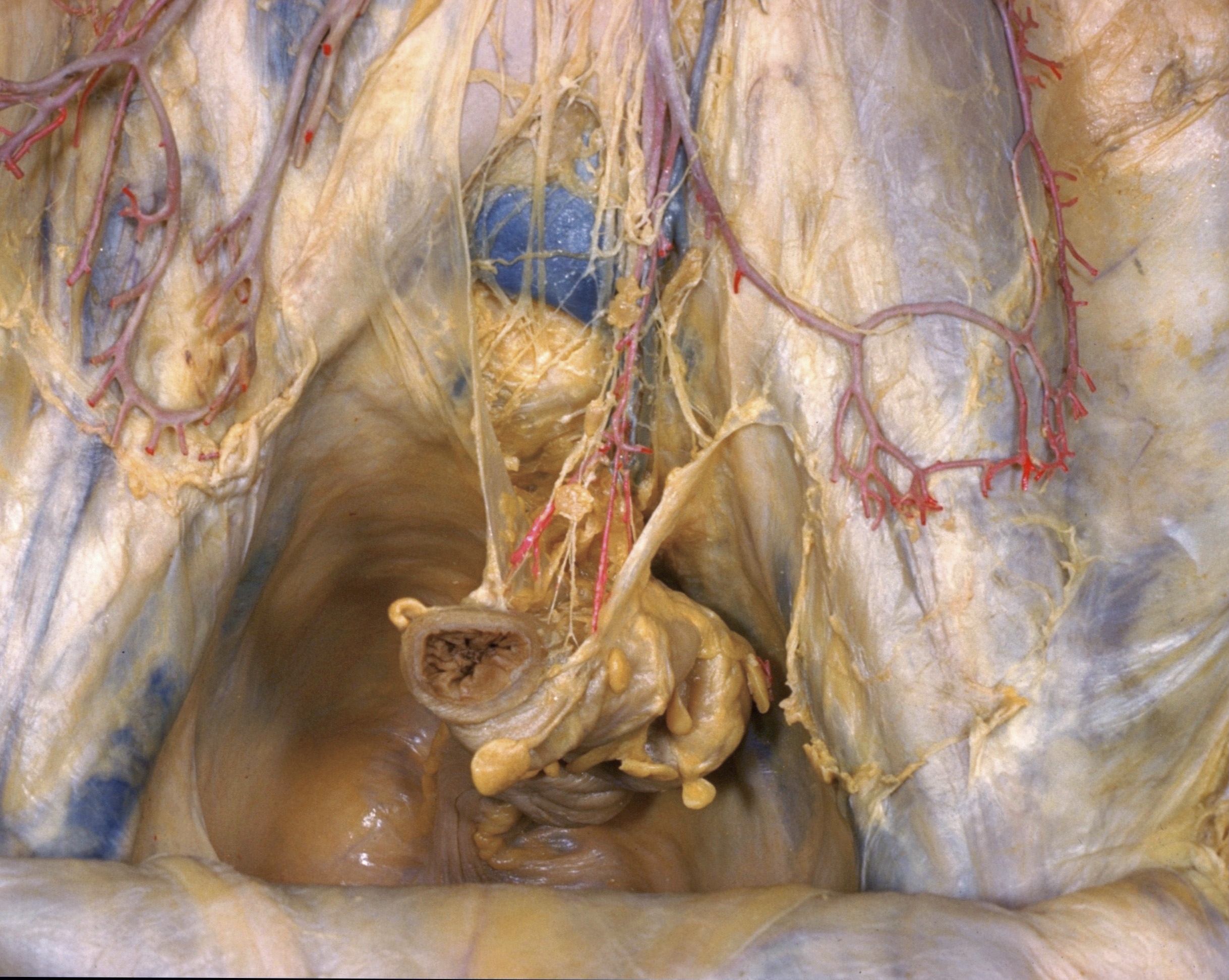 | 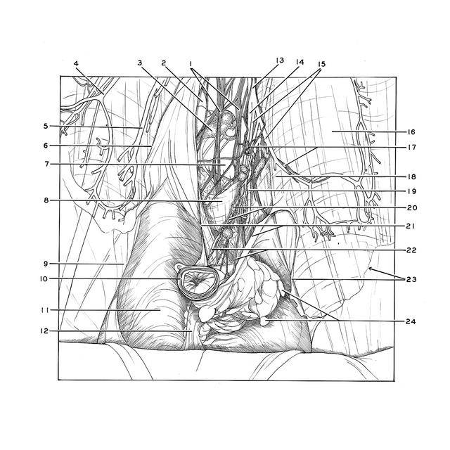 |
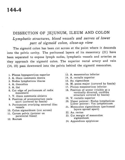 | | Dissection of jejunum, ileum and colon | | Lymphatic structures, blood vessels and nerves of lower part of sigmoid colon, close-up view | | The sigmoid colon has been cut across at the point where it descends into the pelvic cavity. The peritoneal layers of its mesentery (21) have been separated to expose lymph nodes, lymphatic vessels and arteries as they approach the sigmoid colon. The superior rectal artery and vein (14,19) pass downward into the pelvis behind the sigmoid mesocolon. | | 1
.
| Superior hypogastric plexus | | 2
.
| Right common iliac artery | | 3
.
| Iliac lymph node | | 4
.
| Ileocolic artery | | 5
.
| Ileal artery | | 6
.
| Cut edge of peritoneum of root of mesenteries | | 7
.
| Left common iliac vein | | 8
.
| Position of Promontory of sacrum (covered by fascia) | | 9
.
| Peritoneum overlying external iliac vessels | | 10
.
| Sigmoid colon (cut across) | | 11
.
| Pelvic cavity (pointer on pararectal fossa) | | 12
.
| Rectum | | 13
.
| Inferior mesenteric artery | | 14
.
| Superior rectal artery | | 15
.
| Sigmoid arteries | | 16
.
| Psoas major muscle (covered by fascia) | | 17
.
| Inferior mesenteric plexus | | 18
.
| Position of ureter (visible as a vertically directed, cordlike structure covered by fascia) | | 19
.
| Superior rectal vein | | 20
.
| Upper pointer: Lymph node Lower pointer: Lymph vessel | | 21
.
| Mesosigmoid colon (peritoneal layers spread apart) | | 22
.
| Straight arteries | | 23
.
| Cut margin of mesosigmoid colon | | 24
.
| Appendices epiploicae |
|
|


