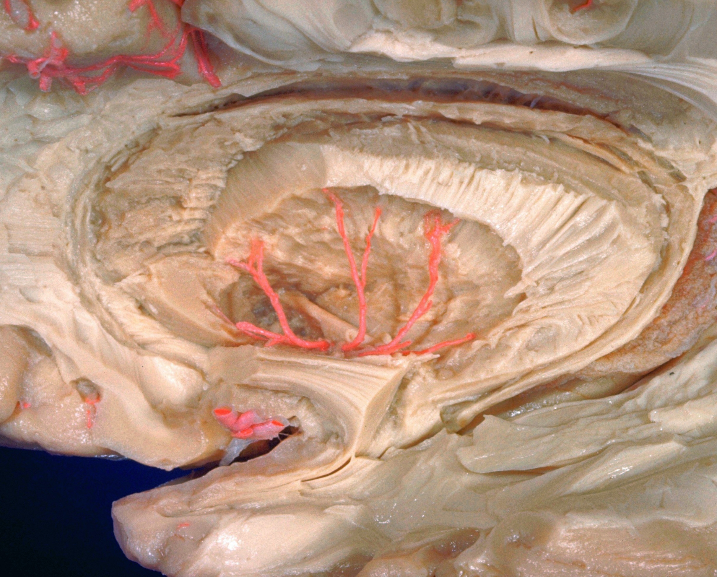
Anterior limb of internal capsule; continuity of head of caudate nucleus with putamen; posterior limb of anterior commissure
For permissions information regarding the use of these images,
please contact Drew Bourn at dbourn@stanford.edu
Image #14-7
Exploration of the brain from its lateral aspect
Anterior limb of internal capsule; continuity of head of caudate nucleus with putamen; posterior limb of anterior commissure
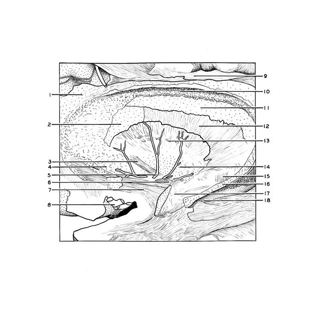
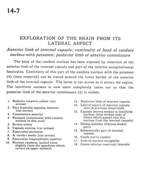
The head of the caudate nucleus has been exposed by resection of the anterior limb of the internal capsule and part of the inferior occipitofrontal fasciculus. Continuity of this part of the caudate nucleus with the putamen (4) (now removed) can be traced around the lower border of the anterior limb of the internal capsule. The latter is cut across as it enters the region. The lentiform nucleus is now more completely taken out so that the posterior limb of the anterior commissure (3) is visible.
1
.
Radiation corpus callosum (cut across)
2
.
Frontal part internal capsule (cut across)
3
.
Anterior commissure
4
.
Putamen (continuous with caudate nucleus in this area)
5
.
Striate artery
6
.
External capsule (cut across)
7
.
Uncinate fasciculus
8
.
Middle cerebral artery (cut across)
9
.
Superior longitudinal fasciculus
10
.
Caudate nucleus (pulled away slightly from the ependyma which covers its upper surface)
11
.
Posterior limb of internal capsule
12
.
Lateral aspect of internal capsule, seen in previous dissections
13
.
Internal capsule medial to lentiform nucleus (note broken ends of fibers which passed into this nucleus from the internal capsule)