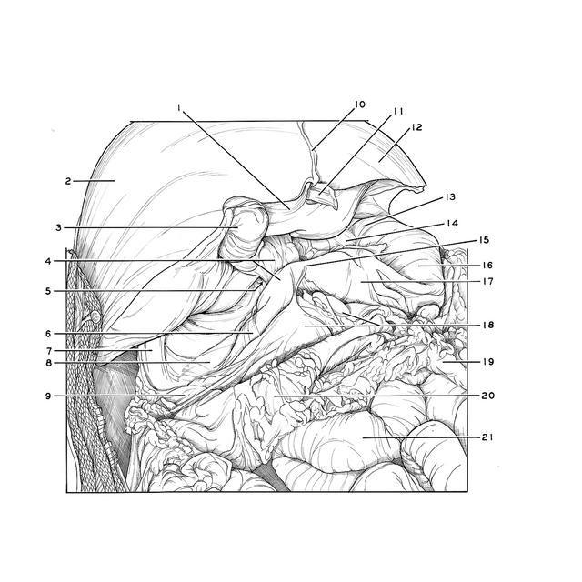
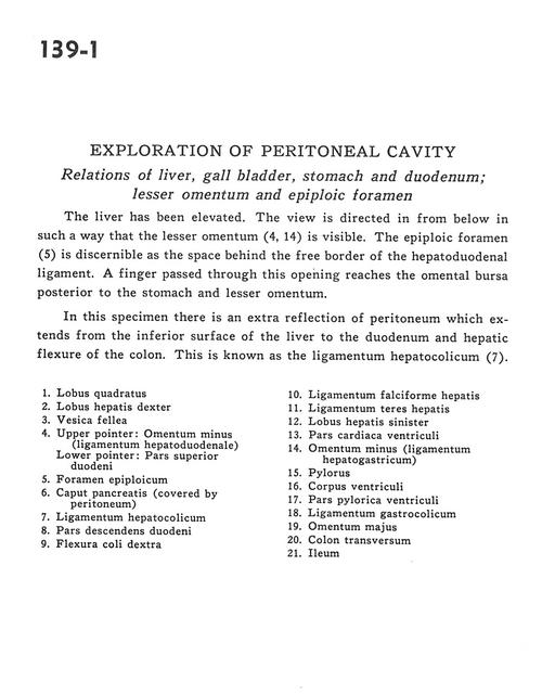
Exploration of peritoneal cavity
Relations of liver, gall bladder, stomach and duodenum; lesser omentum and epiploic foramen
Stanford holds the copyright to the David L. Bassett anatomical images and has assigned
Creative Commons license Attribution-Share Alike 4.0 International to all of the images.
For additional information regarding use and permissions,
please contact Dr. Drew Bourn at dbourn@stanford.edu.
Image #139-1
 |  | ||||||||||||||||||||||||||||||||||||||||||||||
 |
|