X CLOSE

Exploration of peritoneal cavity

Relations of liver, gall bladder, stomach and duodenum; lesser omentum and epiploic foramen

Stanford holds the copyright to the David L. Bassett anatomical images and has assigned Creative Commons license Attribution-Share Alike 4.0 International to all of the images.
For additional information regarding use and permissions, please contact Dr. Drew Bourn at dbourn@stanford.edu.

Image #139-1
Bassett ImageDiagram Image
Legend Image
Exploration of peritoneal cavity
Relations of liver, gall bladder, stomach and duodenum; lesser omentum and epiploic foramen
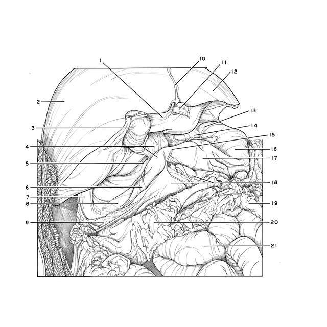
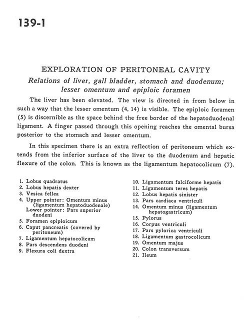
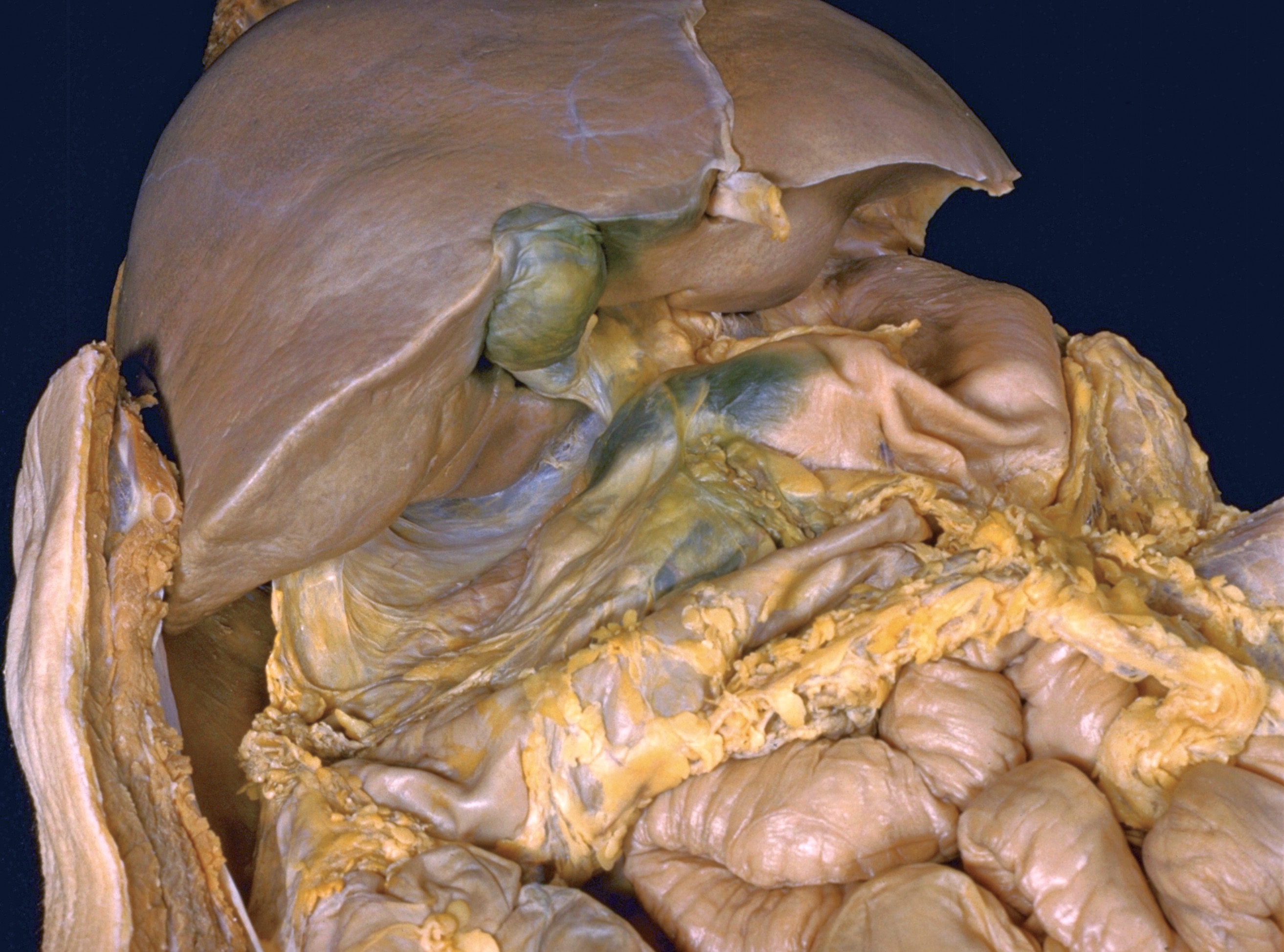
The liver has been eleveated. The view is directed in from below in such a way that the lesser omentum (4,14) is visible. The epiploic foramen (5) is discernable as the space behind the free border of the hepatoduodenal ligament. A finger passed through this opening reaches the omental bursa posterior to the stomach and lesser omentum. In this specimen there is an extra reflection of peritoneum which extends from the inferior surface of the liver to the duodenum and hepatic flexure of colon. This is known as the ligamentum hepatocolicum (7).
1 . Quadrate lobe
2 . Right lobe of liver
3 . Gallbladder
4 . Upper pointer: Lesser omentum (hepatoduodenal ligament) Lower pointer: Superior part of duodenum
5 . Epiploic foramen
6 . Head of pancreas (covered by peritoneum)
7 . Hepatocolic ligament
8 . Descending part of duodenum
9 . Right colic flexure
10 . Falciform ligament of liver
11 . Ligamentum teres (of liver)
12 . Left lobe of liver
13 . Cardiac part of stomach
14 . Lesser omentum (hepatogastric ligament)
15 . Pylorus
16 . Body of stomach
17 . Pyloric part of stomach
18 . Gastrocolic ligament
19 . Greater omentum
20 . Transverse colon
21 . Ileum