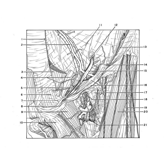
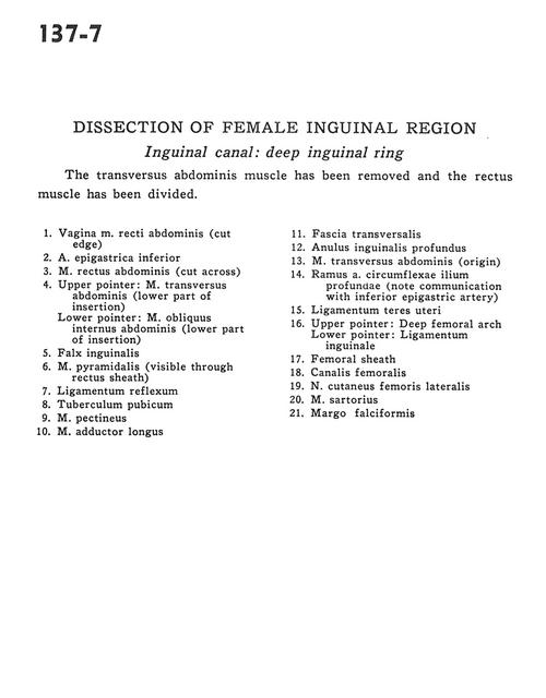
Dissection of female inguinal region
Inguinal canal .
Stanford holds the copyright to the David L. Bassett anatomical images and has assigned
Creative Commons license Attribution-Share
Alike 4.0 International to all of the images.
For additional information regarding use and permissions,
please contact Dr. Drew Bourn at dbourn@stanford.edu.
Image #137-7
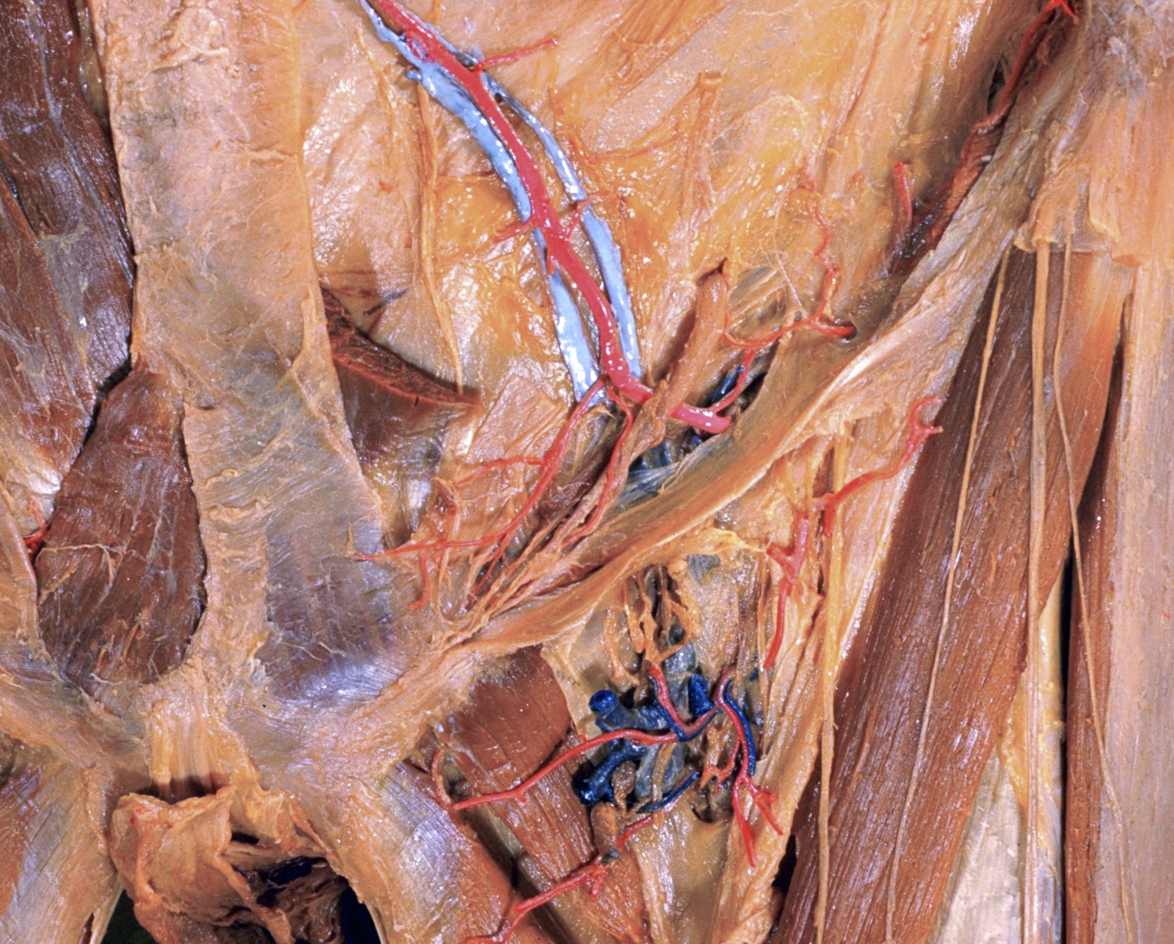
Dissection of female inguinal region
Inguinal canal .
The transversus abdominis muscle has been removed and the rectus muscle has been divided.
- Sheath of rectus abdominis muscle (cut edge)
- Inferior epigastric artery
- Rectus abdominis muscle (cut across)
- Upper pointer: Transversus abdominis muscle (lower part of insertion) Lower pointer: Internal oblique muscle (lower part of insertion)
- Conjoint tendon
- Pyramidalis muscle (visible through rectus sheath)
- Reflected inguinal ligament
- Pubic tubercle
- Pectineus muscle
- Adductor longus muscle
- Transversalis fascia
- Deep inguinal ring
- Transversus abdominis muscle (origin)
- Branch of deep circumflex iliac artery (note communication with inferior epigastric artery)
- Ligamentum teres (of uterus)
- Upper pointer: Deep femoral arch Lower pointer: Inguinal ligament
- Femoral sheath
- Femoral canal
- Lateral femoral cutaneous nerve
- Sartorius muscle
- Falciform margin