X CLOSE

Dissection of female inguinal region

Inguinal canal (continued).

Stanford holds the copyright to the David L. Bassett anatomical images and has assigned Creative Commons license Attribution-Share Alike 4.0 International to all of the images.
For additional information regarding use and permissions, please contact Dr. Drew Bourn at dbourn@stanford.edu.

Image #137-4
Bassett ImageDiagram Image
Legend Image
Dissection of female inguinal region
Inguinal canal (continued).
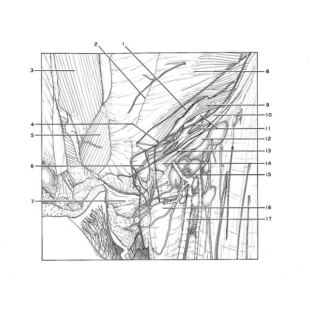
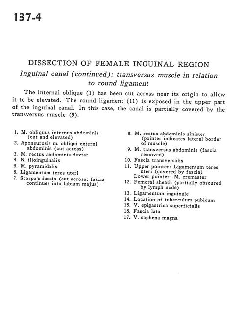
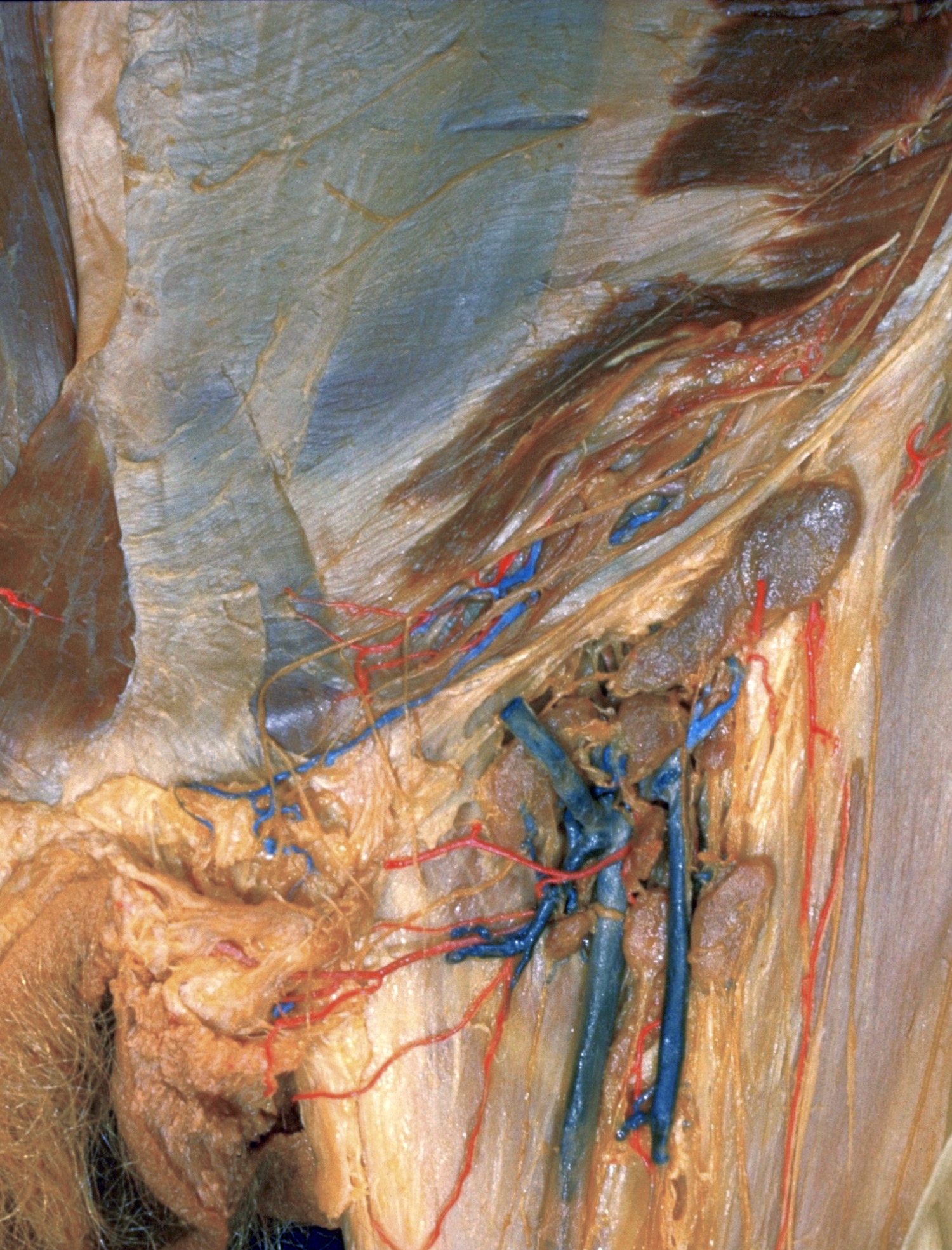
The internal oblique (1) has been cut across near its origin to allow it to be elevated. The round ligament *(11) is exposed in the upper part of the inguinal canal. In this case, the canal is partially covered by the transversus muscle (9).
1 . Internal oblique muscle (cut and elevated)
2 . Aponeurosis External oblique muscle (cut across)
3 . Right rectus abdominis muscle
4 . Ilioinguinal nerve
5 . Pyramidalis muscle
6 . Ligamentum teres (of uterus)
7 . Scarpa's fascia (cut across fascia continues into labium majus)
8 . Left rectus abdominis muscle (pointer indicates lateral border of muscle)
9 . Transversus abdominis muscle (fascia removed)
10 . Transversalis fascia
11 . Upper pointer: Ligamentum teres (of uterus) (covered by fascia) Lower pointer: Cremaster muscle
12 . Femoral sheath (partially obscured by lymph node)
13 . Inguinal ligament
14 . Location of pubic tubercle
15 . Superficial epigastric vein
16 . Fascia lata
17 . Great saphenous vein