X CLOSE

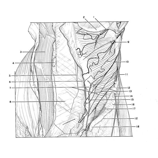
Dissection of anterolateral abdominal wall

Transversus abdominis muscle, left anterolateral view

Stanford holds the copyright to the David L. Bassett anatomical images and has assigned Creative Commons license Attribution-Share Alike 4.0 International to all of the images.
For additional information regarding use and permissions, please contact Dr. Drew Bourn at dbourn@stanford.edu.

Image #134-3
Bassett ImageDiagram Image
Legend Image
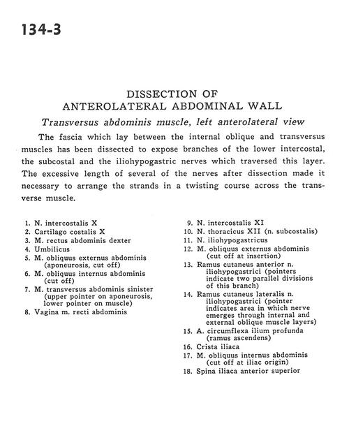
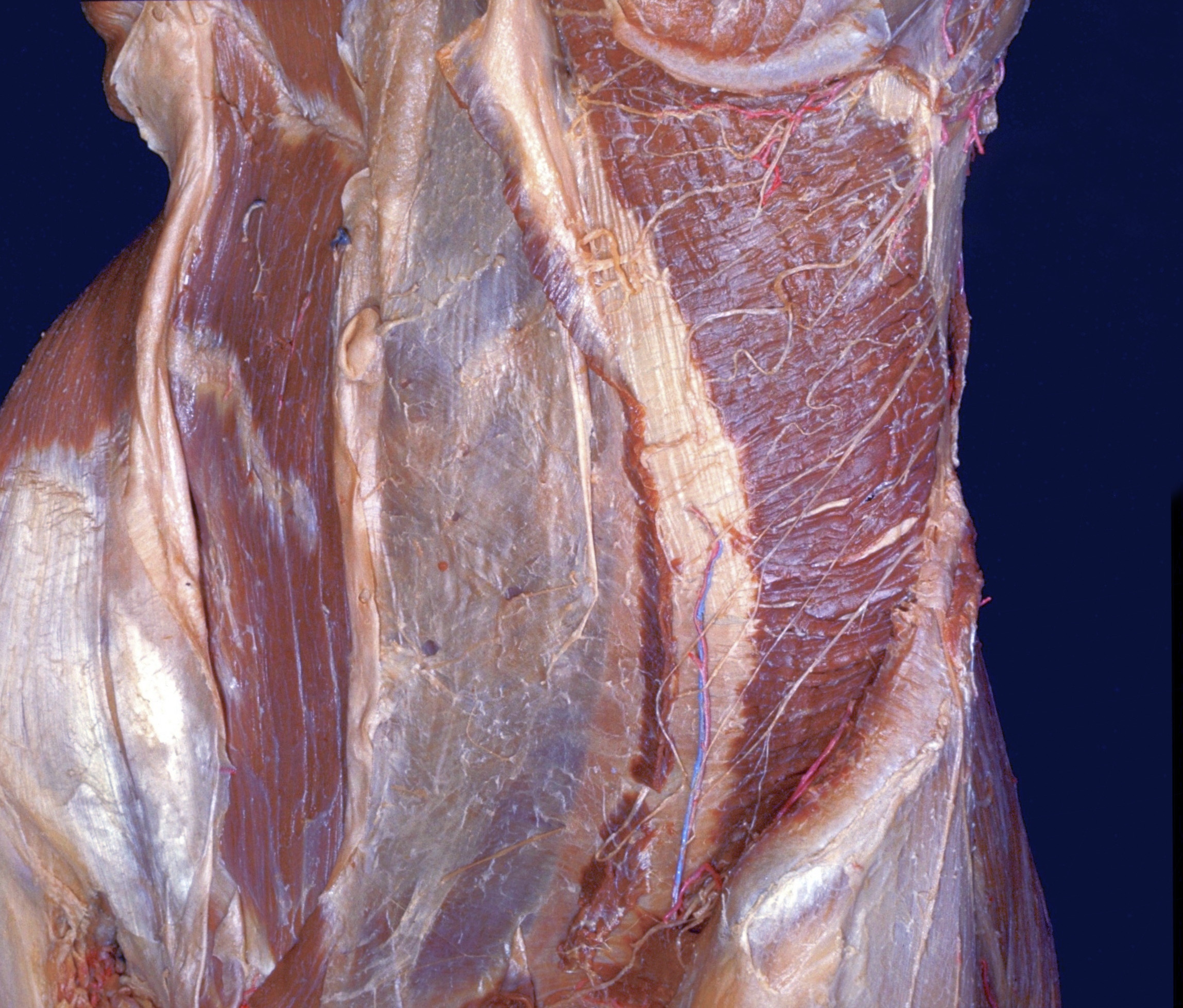
Dissection of anterolateral abdominal wall
Transversus abdominis muscle, left anterolateral view
The fascia which lay between the internal oblique and transversus muscles has been dissected to expose branches of the lower intercostal, the subcostal and the iliohypogastric nerves which traversed this layer. The excessive length of several of the nerves after dissection made it necessary to arrange the strands in a twisting course across the transverse muscle.
1 . Intercostal nerve X
2 . Costal cartilage X
3 . Right rectus abdominis muscle
4 . Umbilicus
5 . External oblique muscle (aponeurosis, cut off)
6 . Internal oblique muscle (cut off)
7 . Left transversus abdominis muscle (upper pointer on aponeurosis, lower pointer on muscle)
8 . Sheath of rectus abdominis muscle
9 . Intercostal nerve XI
10 . Thoracic nerve XII (subcostal nerve)
11 . Iliohypogastric nerve
12 . External oblique muscle (cut off at insertion)
13 . Anterior cutaneous branch iliohypogastric nerve (pointers indicate two parallel divisions of this branch)
14 . Lateral cutaneous branch iliohypogastric nerve (pointer indicates area in which nerve emerges through internal and external oblique muscle layers)
15 . Deep circumflex iliac artery (ascending branch)
16 . Iliac crest
17 . Internal oblique muscle (cut off at iliac origin)
18 . Anterior superior iliac spine