X CLOSE

Dissection of anterolateral abdominal wall

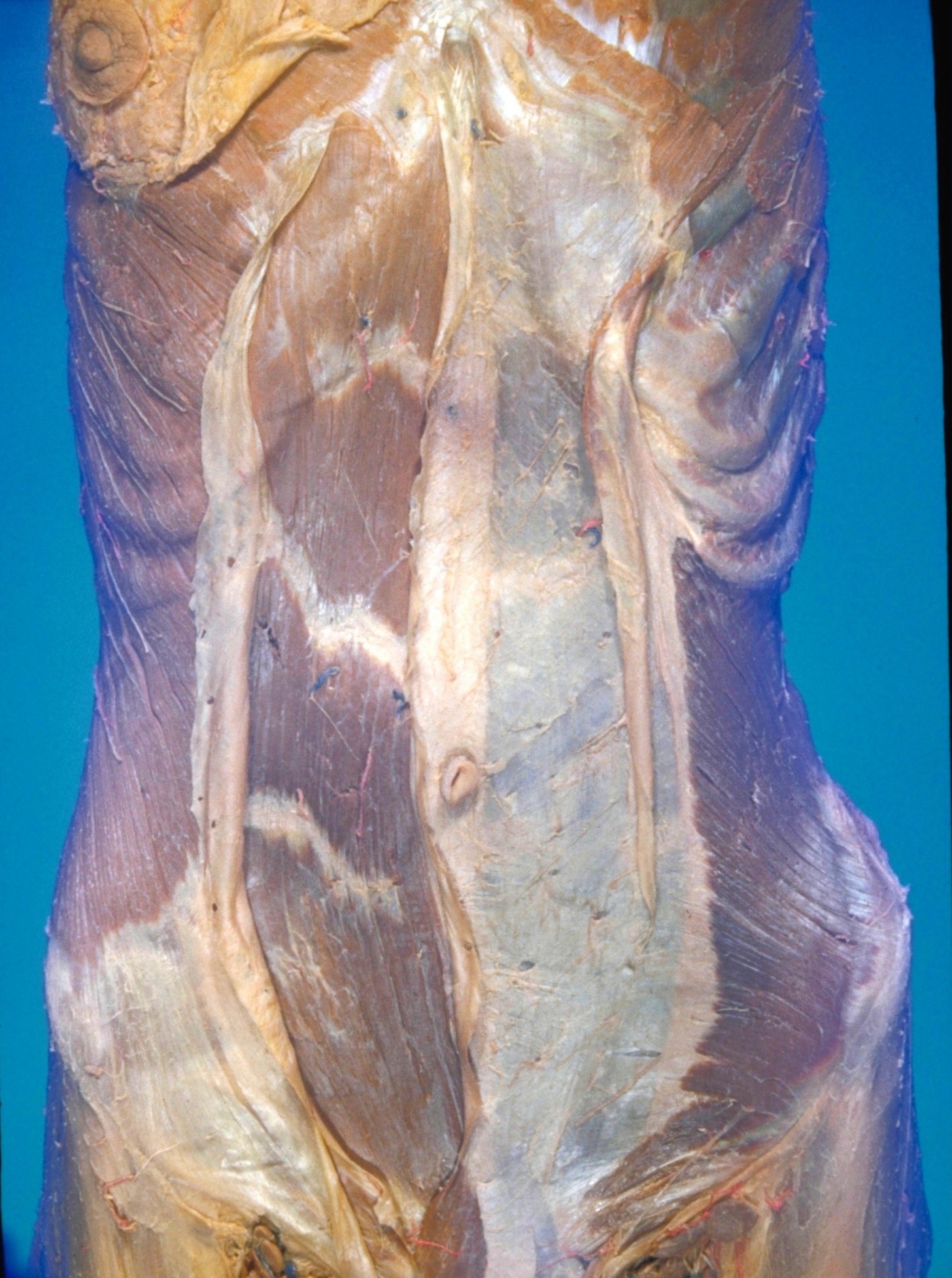
General anterior view of external oblique, internal oblique and rectus abdominis muscles

For permissions information regarding the use of these images, please contact Drew Bourn at dbourn@stanford.edu

Image #133-6
Bassett ImageDiagram Image
Legend Image
Dissection of anterolateral abdominal wall
General anterior view of external oblique, internal oblique and rectus abdominis muscles
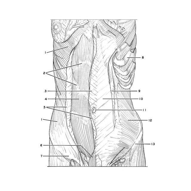
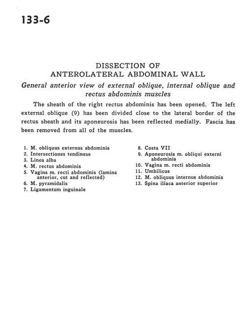
The sheath of the right rectus abdominis has been opened. The left external oblique (9) has been divided close to the lateral border of the rectus sheath and its aponeurosis has been reflected medially. Fascia has been removed from all of the muscles.
1 . External oblique muscle
2 . Tendinous inscriptions
3 . Linea alba
4 . Rectus abdominis muscle
5 . Sheath of rectus abdominis muscle (anterior layer, cut and reflected)
6 . Pyramidalis muscle
7 . Inguinal ligament
8 . Rib VII
9 . Aponeurosis external oblique muscle
10 . Sheath of rectus abdominis muscle
11 . Umbilicus
12 . Internal oblique muscle
13 . Anterior superior iliac spine