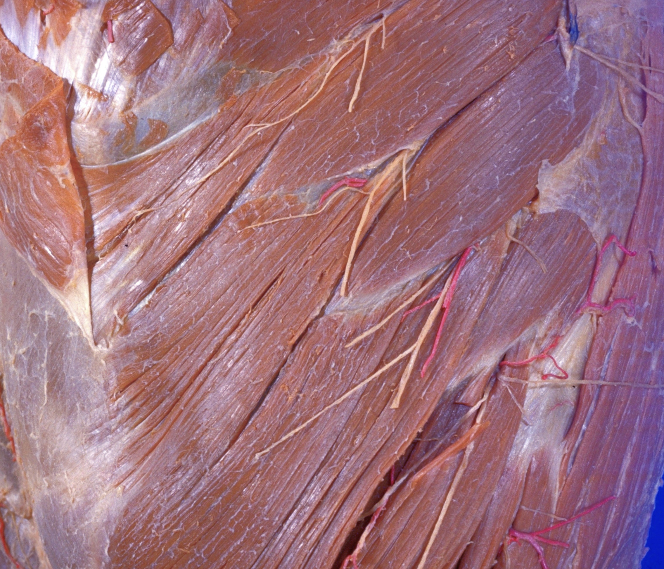
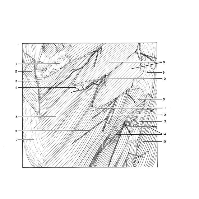
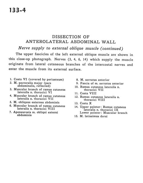
Dissection of anterolateral abdominal wall
Nerve supply to external oblique muscle (continued)
Stanford holds the copyright to the David L. Bassett anatomical images and has assigned
Creative Commons license Attribution-Share Alike 4.0 International to all of the images.
For additional information regarding use and permissions,
please contact Dr. Drew Bourn at dbourn@stanford.edu.
Image #133-4
 |  | ||||||||||||||||||||||||||||||||
 |
|