Exploration of the brain from its lateral aspect
Internal capsule and superior longitudinal fasciculus
Stanford holds the copyright to the David L. Bassett anatomical images and has assigned
Creative Commons license Attribution-Share
Alike 4.0 International to all of the images.
For additional information regarding use and permissions,
please contact Dr. Drew Bourn at dbourn@stanford.edu.
Image #13-6
Exploration of the brain from its lateral aspect
Internal capsule and superior longitudinal fasciculus
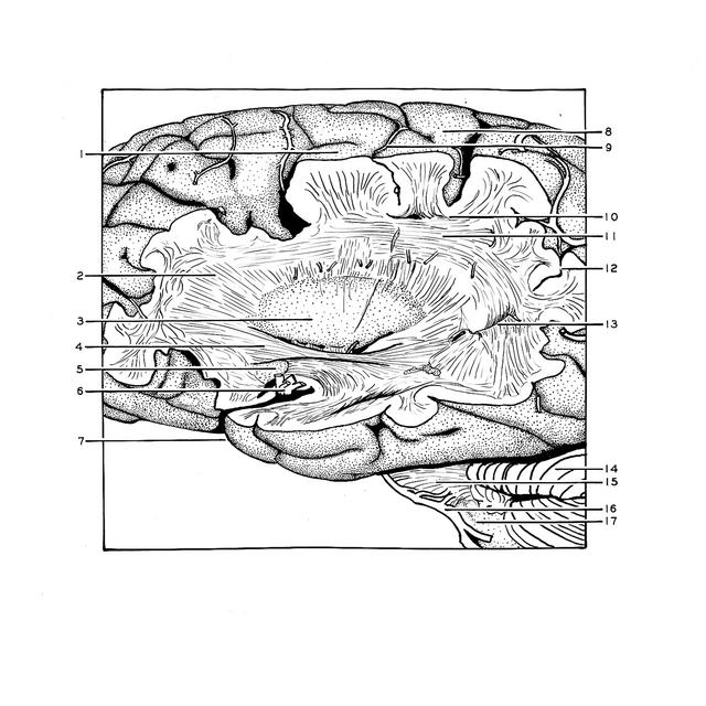
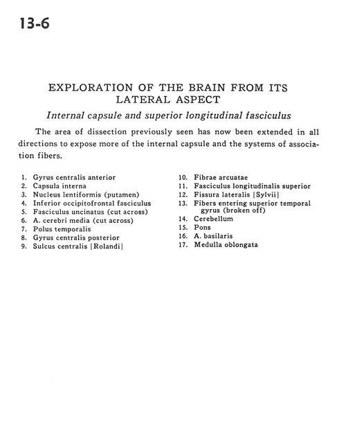
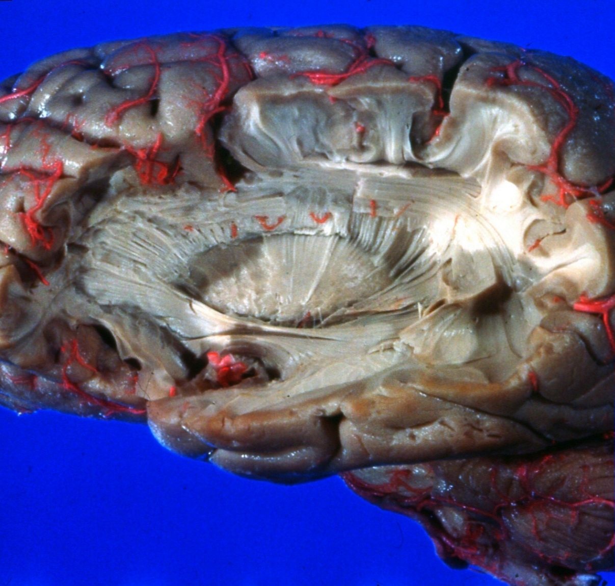
The areas of dissection previously seen has now been extended in all directions to expose more of the internal capsule and the systems of association fibers.
- Precentral gyrus
- Internal capsule
- Lentiform nucleus (putamen)
- Inferior occipitofrontal fasciculus
- Uncinate fasciculus (cut across)
- Middle cerebral artery (cut across)
- Temporal pole
- Postcentral gyrus
- Central sulcus (Rolandic)
- Arcuate fibers
- Superior longitudinal fasciculus
- Lateral fissure (Sylvian)
- Fibers entering superior temporal gyrus (broken off)
- Cerebellum
- Pons
- Basilar artery
- Medulla oblongata