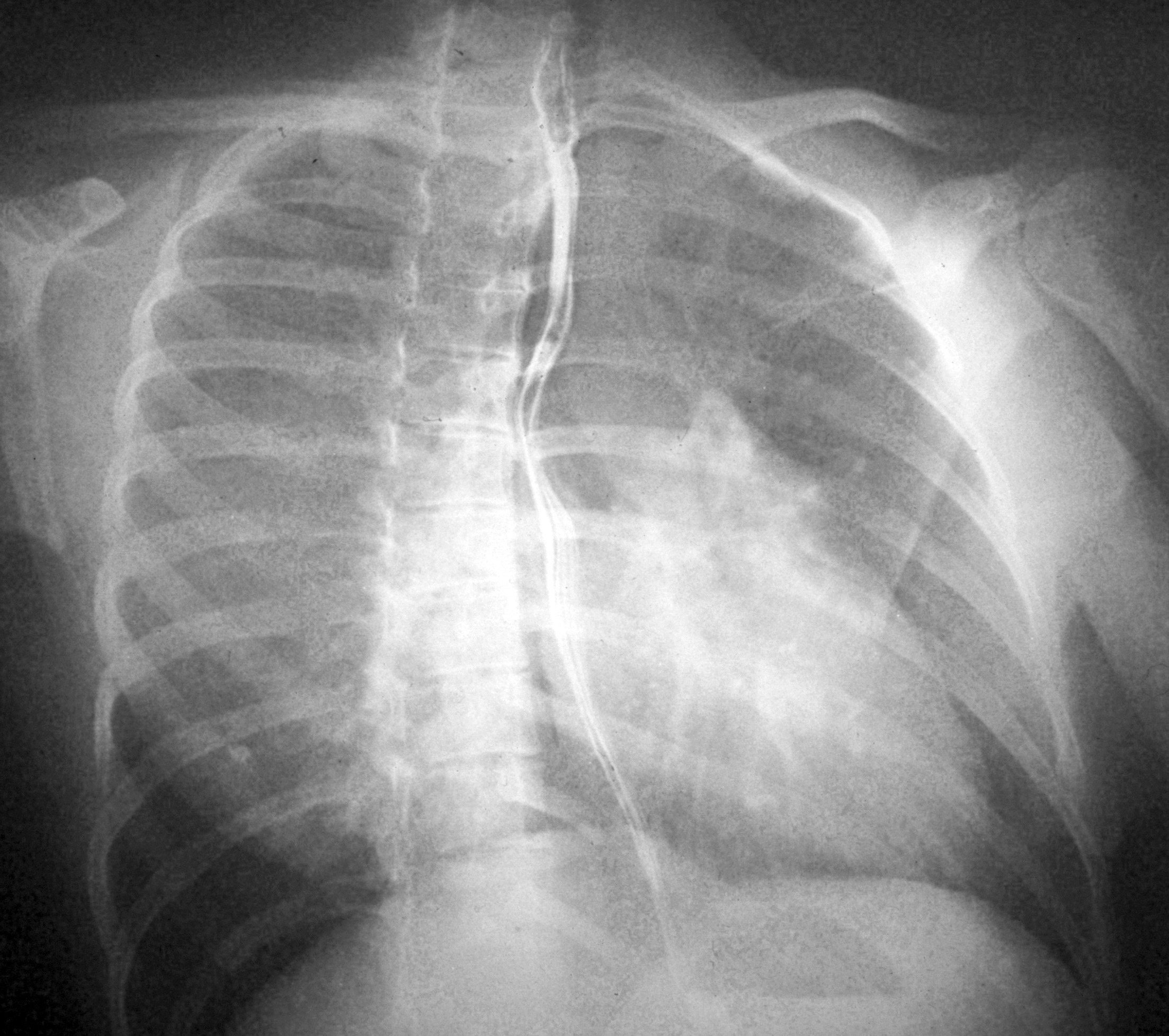
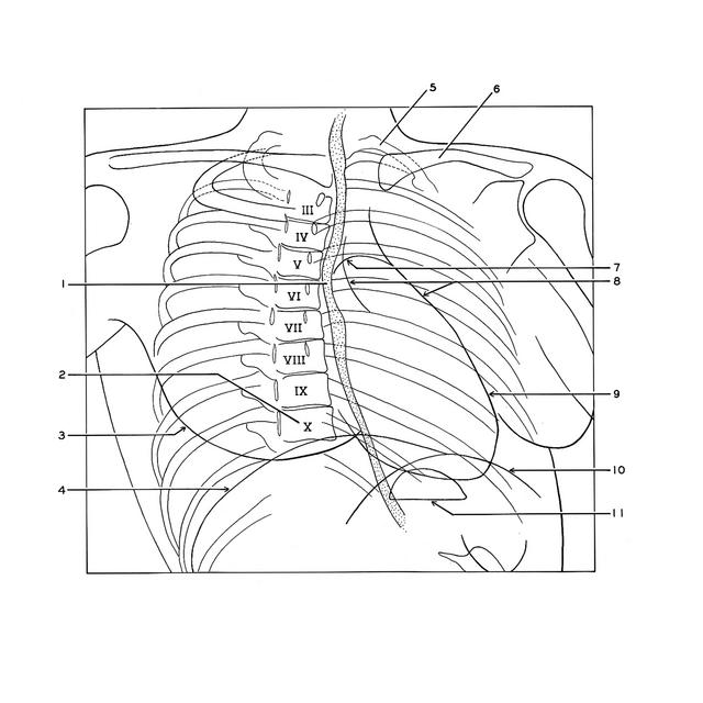
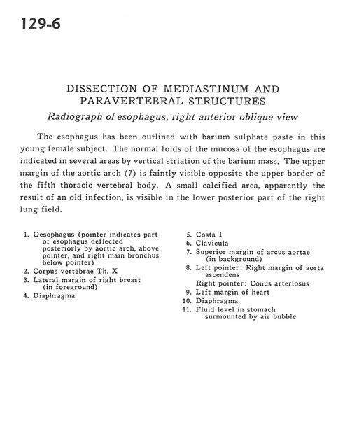
Dissection of mediastinum and paravertebral structures
Radiograph of esophagus, right anterior oblique view
Stanford holds the copyright to the David L. Bassett anatomical images and has assigned
Creative Commons license Attribution-Share Alike 4.0 International to all of the images.
For additional information regarding use and permissions,
please contact Dr. Drew Bourn at dbourn@stanford.edu.
Image #129-6
 |  | ||||||||||||||||||||||||||
 |
|