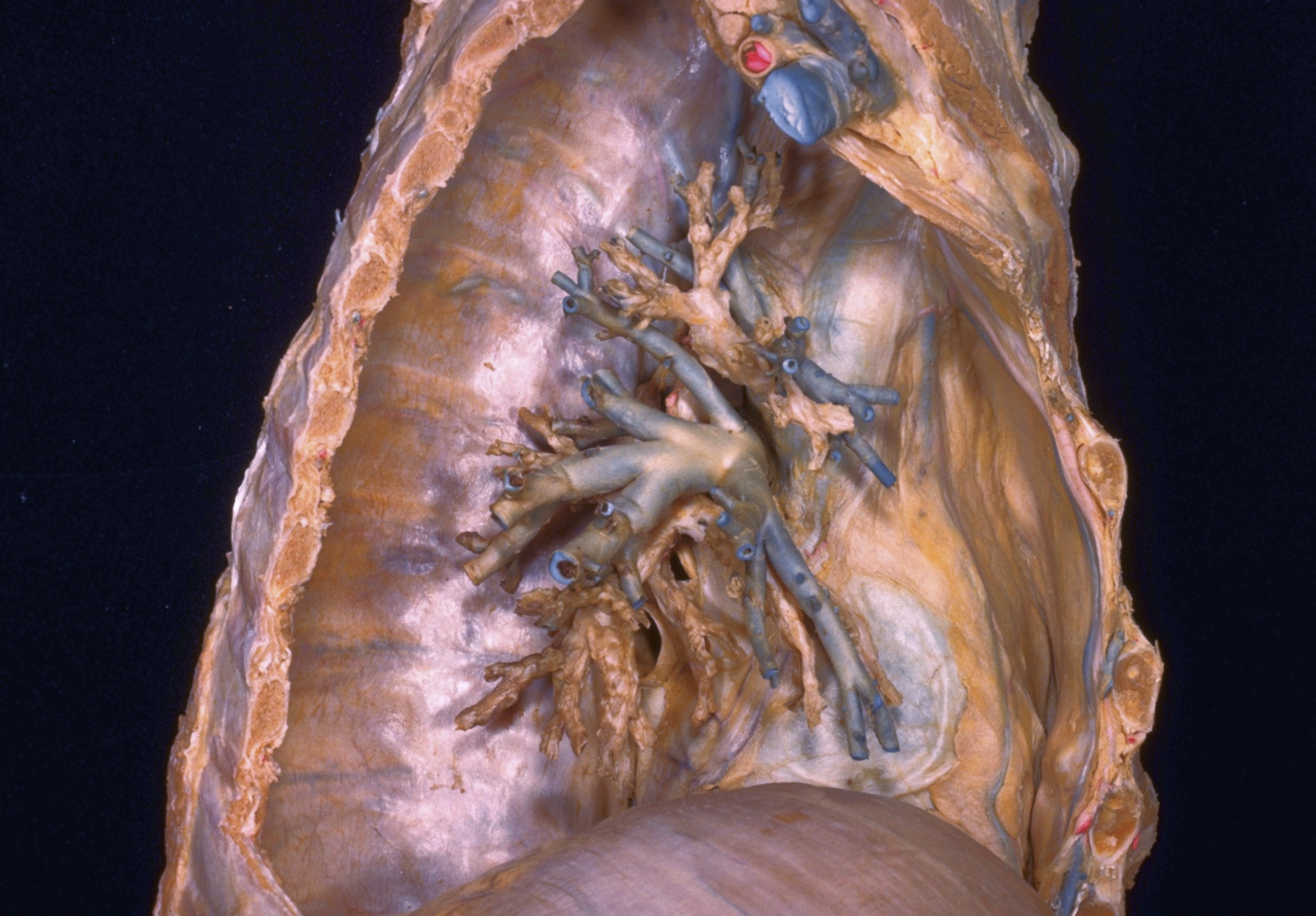
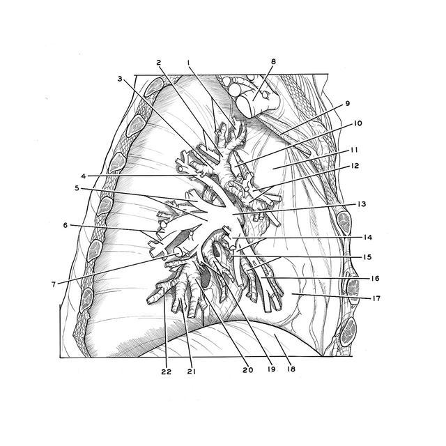
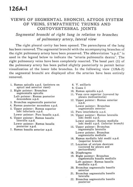
Views of segmental bronchi, azygos system of veins, sympathetic trunks and costovertebral joints
Segmental bronchi of right lung in relation to branches of pulmonary artery, lateral view
Stanford holds the copyright to the David L. Bassett anatomical images and has assigned
Creative Commons license Attribution-Share Alike 4.0 International to all of the images.
For additional information regarding use and permissions,
please contact Dr. Drew Bourn at dbourn@stanford.edu.
Image #126A-1
 |  | ||||||||||||||||||||||||||||||||||||||||||||||||||
 |
|