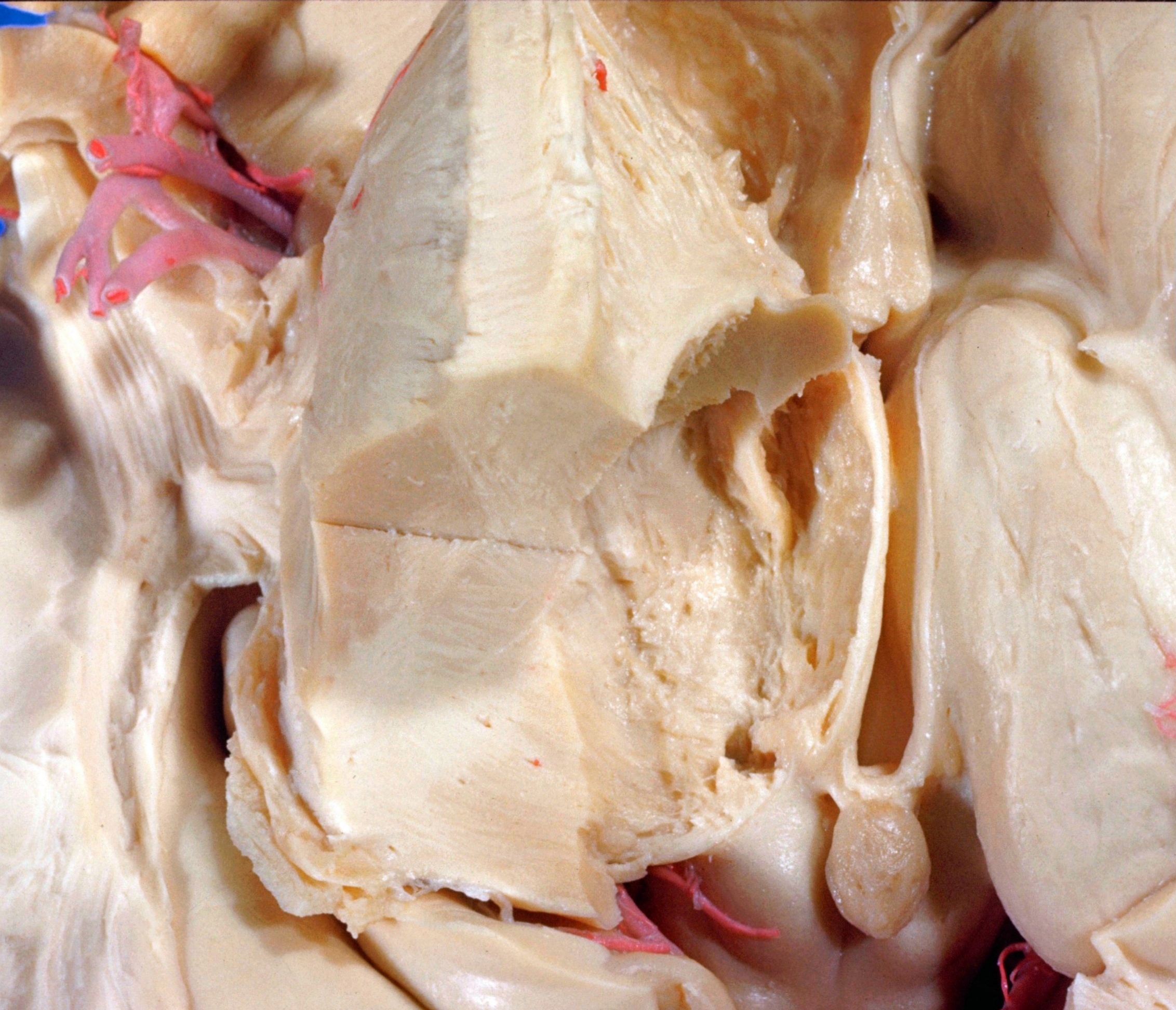
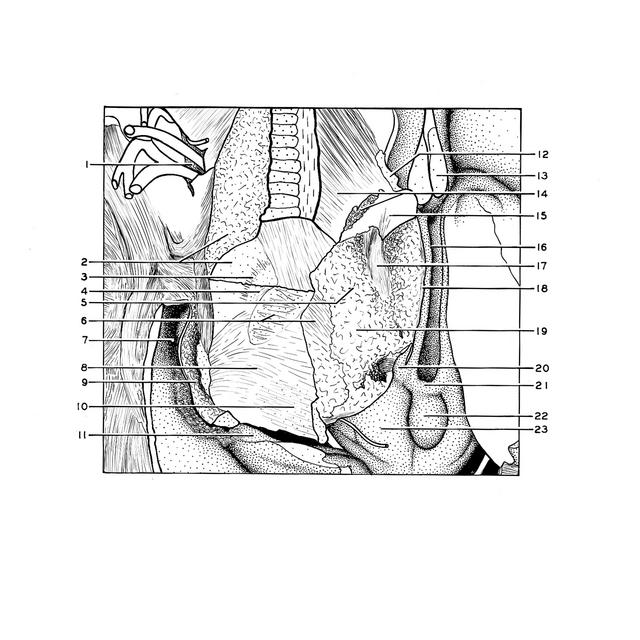
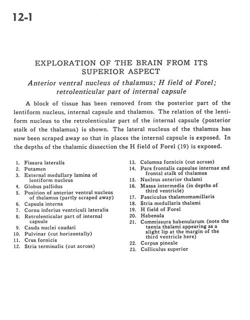
Exploration of the brain from its superior aspect
Anterior ventral nucleus of thalamus; H field of Forel; retrolenticular part of internal capsule
Stanford holds the copyright to the David L. Bassett anatomical images and has assigned
Creative Commons license Attribution-Share Alike 4.0 International to all of the images.
For additional information regarding use and permissions,
please contact Dr. Drew Bourn at dbourn@stanford.edu.
Image #12-1
 |  | ||||||||||||||||||||||||||||||||||||||||||||||||||
 |
|