X CLOSE

Detailed dissection of heart

Posterior surface of heart, epicardium intact

Stanford holds the copyright to the David L. Bassett anatomical images and has assigned Creative Commons license Attribution-Share Alike 4.0 International to all of the images.
For additional information regarding use and permissions, please contact Dr. Drew Bourn at dbourn@stanford.edu.

Image #119-5
Bassett ImageDiagram Image
Legend Image
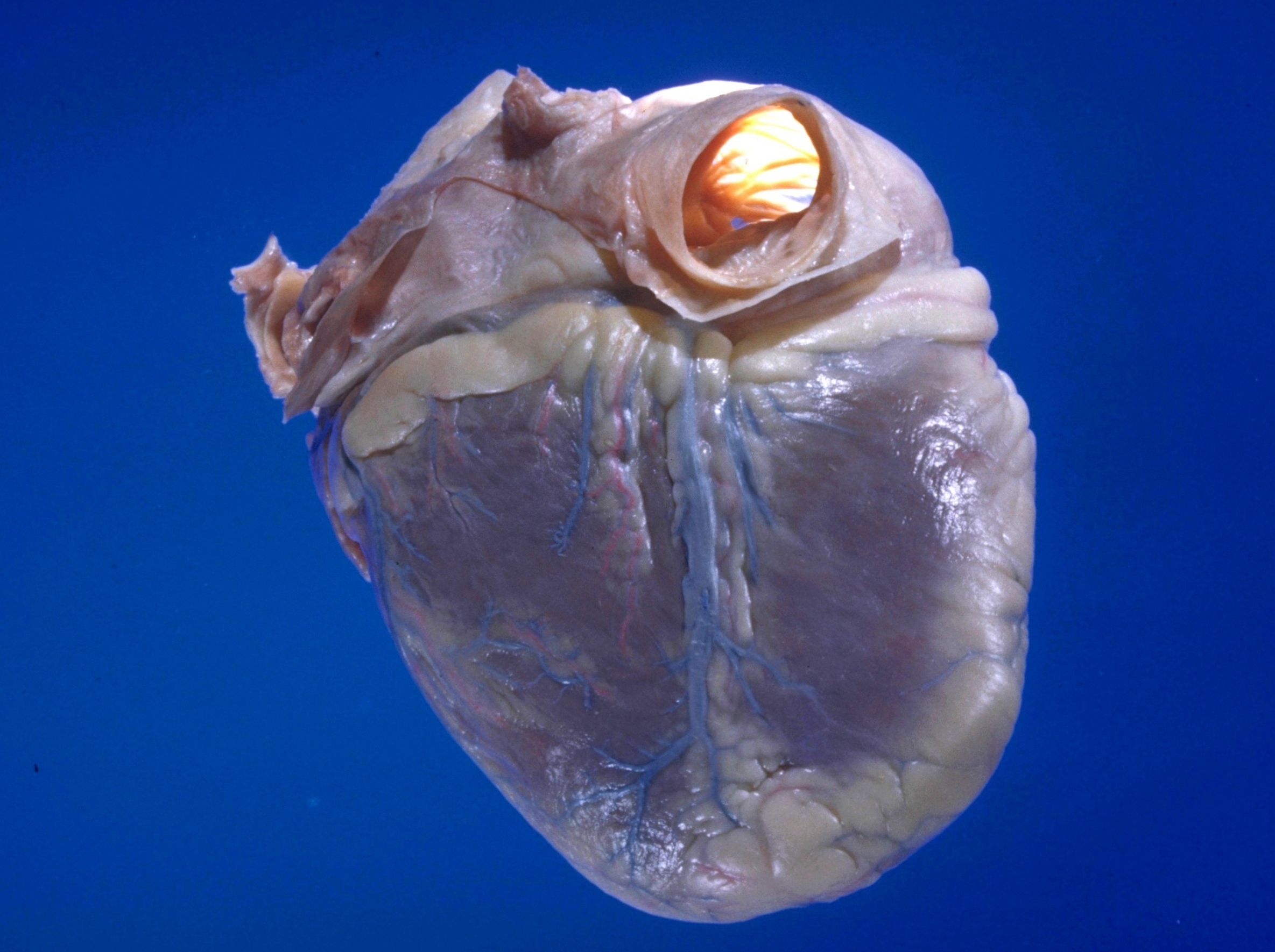
Detailed dissection of heart
Posterior surface of heart, epicardium intact
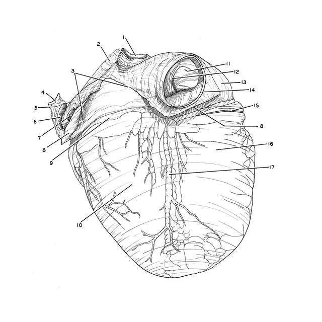
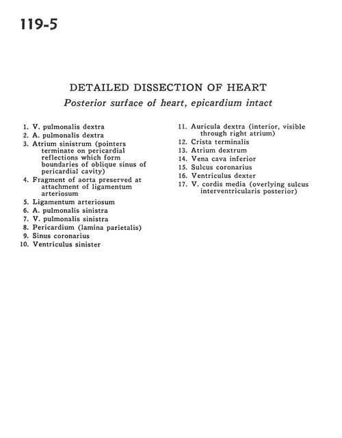
1 . Right pulmonary vein
2 . Right pulmonary artery
3 . Left atrium (pointers terminate on pericardial reflections which form boundaries of oblique sinus of pericardial cavity)
4 . Fragment of aorta preserved at attachment of ligamentum arteriosum
5 . Ligamentum arteriosum
6 . Left pulmonary artery
7 . Left pulmonary vein
8 . Pericardium (parietal side)
9 . Coronary sinus
10 . Left ventricle
11 . Right auricle (interior, visible through right atrium)
12 . Crista terminalis
13 . Right atrium
14 . Inferior vena cava
15 . Coronary sulcus
16 . Right ventricle
17 . Middle cardiac vein (overlying posterior interventricular sulcus)