Transverse section of middle third of hand, partially dissected
For permissions information regarding the use of these images,
please contact Drew Bourn at dbourn@stanford.edu
Image #112-5
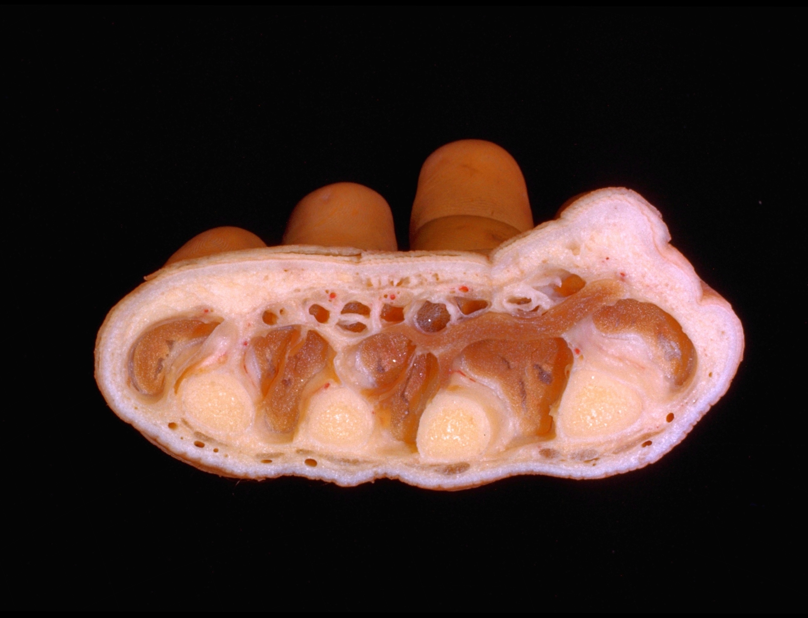
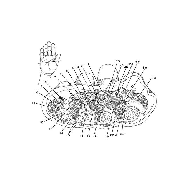
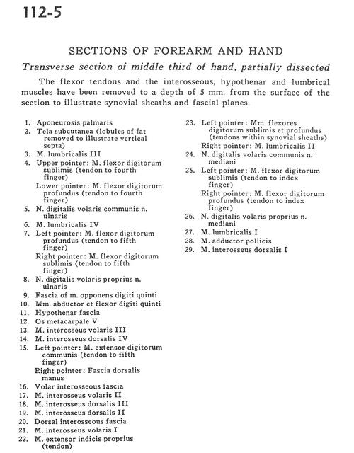
Sections of forearm and hand
Transverse section of middle third of hand, partially dissected
The flexor tendons and the interosseous, hypothenar and lumbrical muscles have been removed to a depth of 5 mm. from the surface of the section to illustrate synovial sheaths and fascial planes.
1
.
Palmar aponeurosis
2
.
Superficial fascia (lobules of fat removed to illustrate vertical septa)
3
.
Lumbrical muscle III
4
.
Upper pointer: Flexor digitorum superficialis (tendon to fourth finger) Lower pointer: Flexor digitorum profundus muscle (tendon to fourth finger)
5
.
Common palmar digital nerve of ulnar nerve
6
.
Lumbrical muscle IV
7
.
Left pointer: Flexor digitorum profundus muscle (tendon to fifth finger) Right pointer: Flexor digitorum superficialis muscle (tendon to fifth finger)
8
.
Proper palmar digital nerve of ulnar nerve
9
.
Fascia of opponens digiti minimi muscle
10
.
Abductor and flexor digiti minimi muscles
11
.
Hypothenar fascia
12
.
Metacarpal V
13
.
Anterior interosseous muscle III
14
.
Dorsal interosseous muscle IV
15
.
Left pointer: Common extensor digitorum muscle (tendon to fifth finger) Right pointer: Dorsal fascial of the hand
16
.
Anterior interosseous fascia
17
.
Anterior interosseous muscle II
18
.
Dorsal interosseous muscle III
19
.
Dorsal interosseous muscle II
20
.
Dorsal interosseous fascia
21
.
Anterior interosseous muscle I
22
.
Extensor indicis muscle (tendon)
23
.
Left pointer: Flexor digitorum superficialis and profundus muscles (tendons within synovial sheaths) Right pointer: Lumbrical muscle II
24
.
Common palmar digital nerve of median nerve
25
.
Left pointer: Flexor digitorum superficialis (tendon to index finger) Right pointer: Flexor digitorum profundus muscle (tendon to index finger)