Joints of right wrist and hand
Articular cavities, anterior view
Stanford holds the copyright to the David L. Bassett anatomical images and has assigned
Creative Commons license Attribution-Share
Alike 4.0 International to all of the images.
For additional information regarding use and permissions,
please contact Dr. Drew Bourn at dbourn@stanford.edu.
Image #110-6
Joints of right wrist and hand
Articular cavities, anterior view
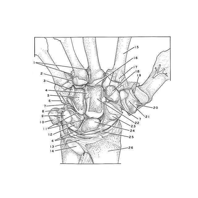
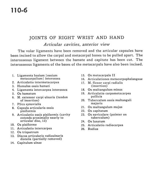
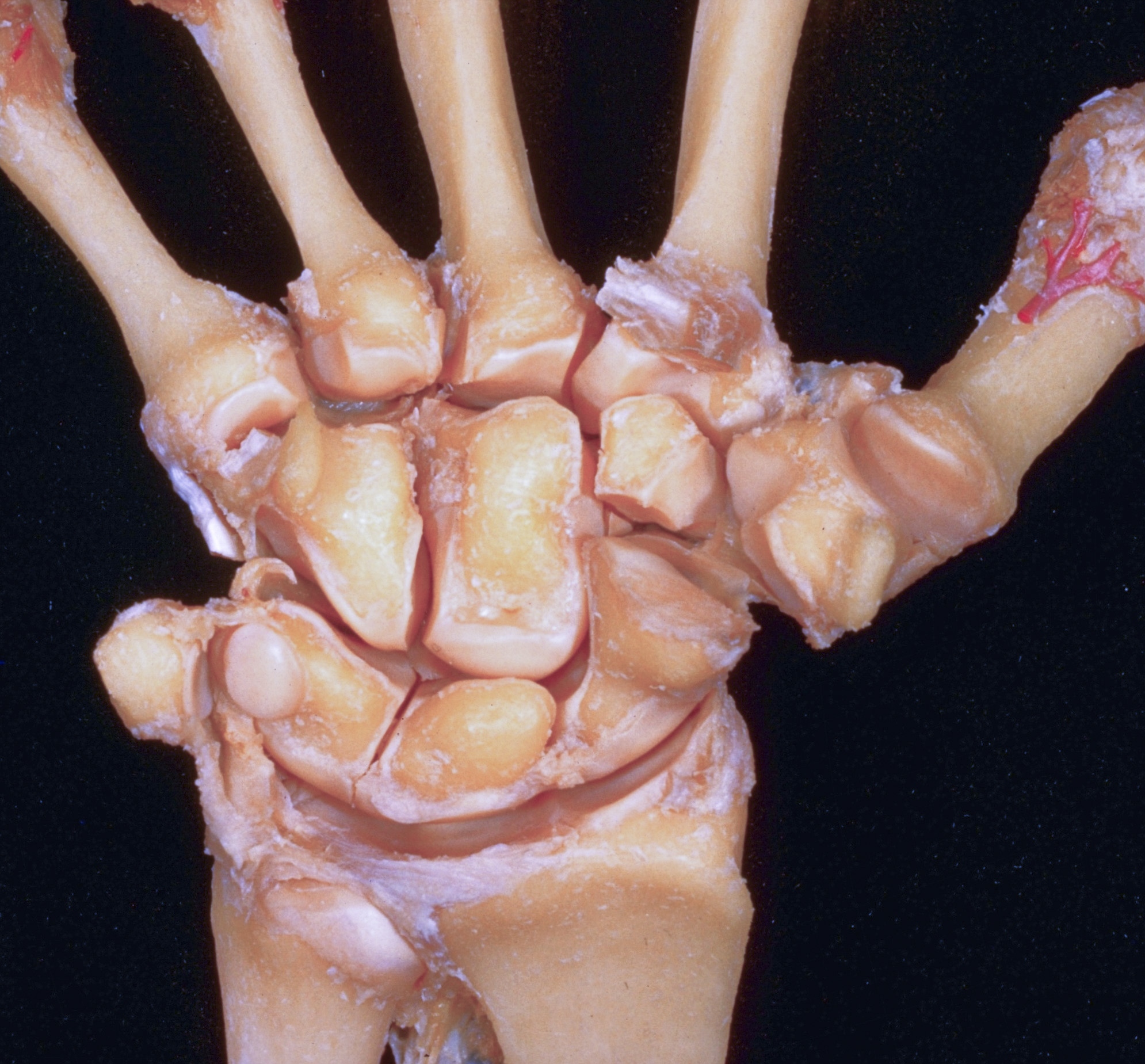
The volar ligaments have been removed and the articular capsules have been incised to allow the carpal and metacarpal bones to be pulled apart. The interosseous ligament between the hamate and capitate has been cut. The interosseous ligaments of the bases of the metacarpals have also been incised.
- Palmar metacarpal ligaments
- Intermetacarpal joint
- Hamulus of hamate bone
- Interosseous intercarpal ligament
- Hamate bone
- Extensor carpi ulnaris muscle (tendon of insertion)
- Synovial fold
- Joint capsule of pisiform bone
- Joint of pisiform bone (cavity extends proximally nearly to articular disc, 13)
- Pisiform bone
- Intercarpal joint
- Triquetral bone
- Distal radioulnar articular disc (partially removed)
- Head of ulna
- Metacarpal II
- Metacarpophalangeal joints
- Flexor carpi radialis muscle (insertion)
- Trapezoid bone
- Metacarpotrapezial joint
- Tubercle of trapezium bone
- Trapezium bone
- Capitate bone
- Scaphoid bone (pointer on tubercle)
- Lunate bone
- Radiocarpal joint
- Radius