Exploration of the brain from its superior aspect
Posterior temporal branch of middle cerebral artery in superior temporal sulcus
Stanford holds the copyright to the David L. Bassett anatomical images and has assigned
Creative Commons license Attribution-Share
Alike 4.0 International to all of the images.
For additional information regarding use and permissions,
please contact Dr. Drew Bourn at dbourn@stanford.edu.
Image #11-2
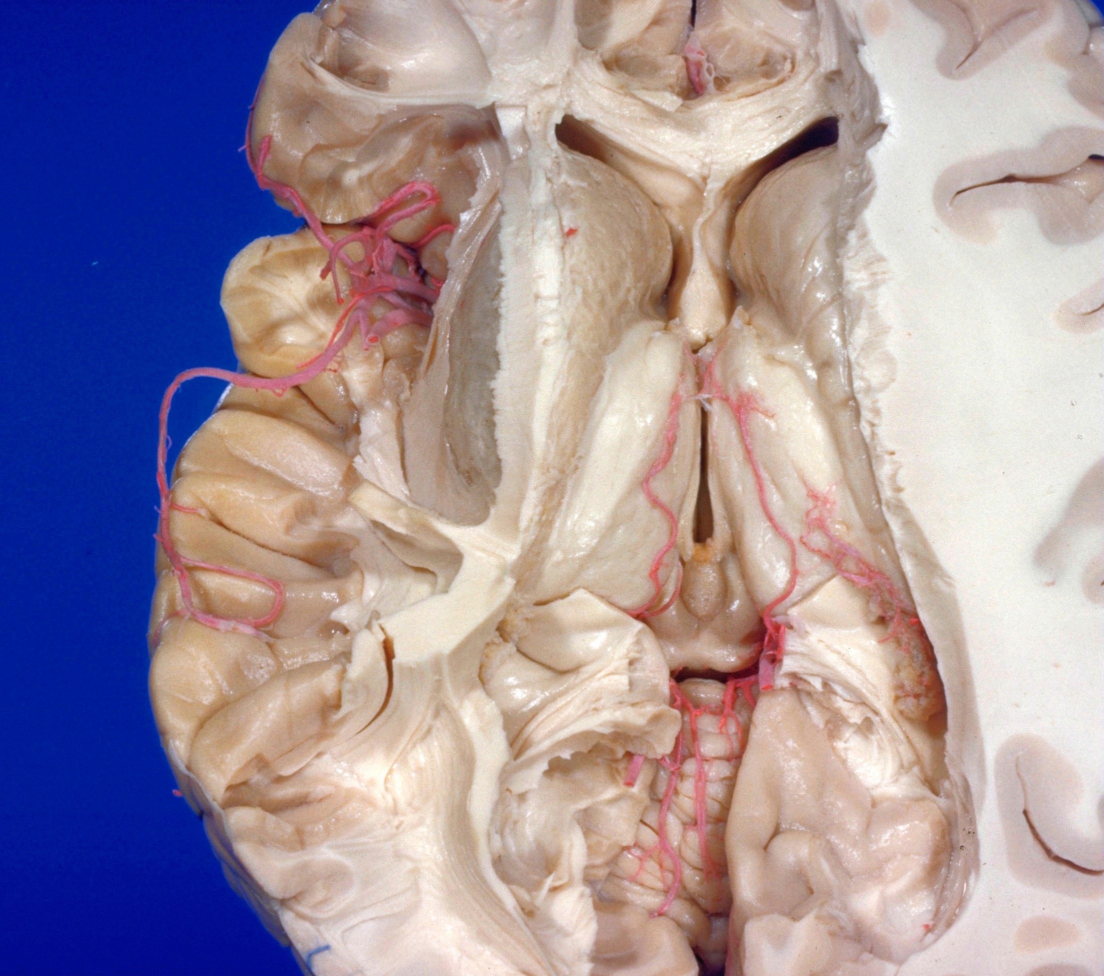
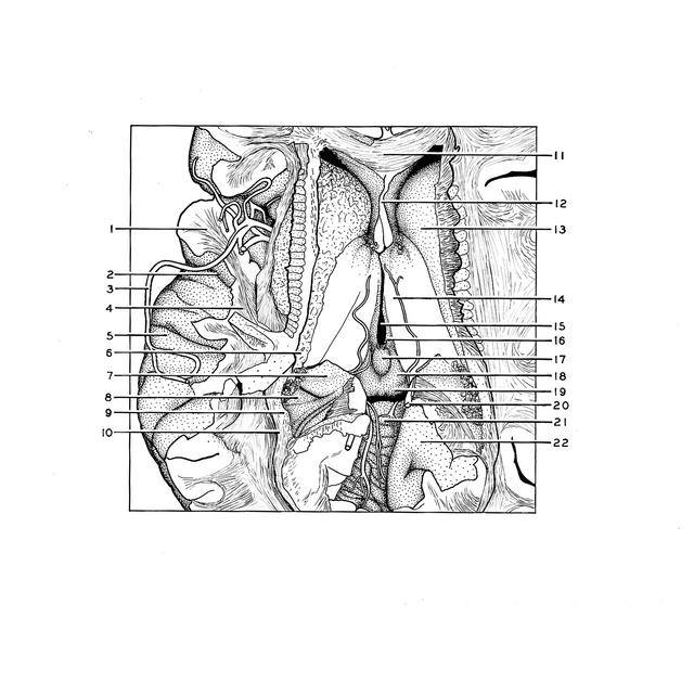
Exploration of the brain from its superior aspect
Posterior temporal branch of middle cerebral artery in superior temporal sulcus
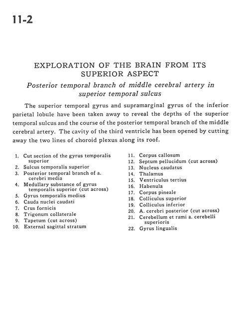
The superior temporal gyrus and supramarginal gyrus of the inferior parietal lobule have been taken away to reveal the depths of the superior temporal sulcus and the course of the posterior temporal branch of the middle cerebral artery. The cavity of the third ventricle has been opened by cutting away the two lines of choroid plexus along its roof.
- Cut section of the superior temporal gyrus
- Superior temporal sulcus
- Posterior temporal branch of middle cerebral artery
- Medullary substance of superior temporal gyrus (cut across)
- Medial temporal gyrus
- Caudate nucleus (tail)
- Fornix (crus)
- Collateral trigone
- Tapetum (cut across)
- External sagittal stratum
- Corpus callosum
- Septum pellucidum (cut across)
- Caudate nucleus
- Thalamus
- Third ventricle
- Habenula
- Pineal body
- Superior colliculus
- Inferior colliculus
- Posterior cerebral artery (cut across)
- Cerebellum and branch superior cerebellar artery
- Lingual gyrus