Volar aspect of right hand
Superficial structures of palm, close-up view
Stanford holds the copyright to the David L. Bassett anatomical images and has assigned
Creative Commons license Attribution-Share
Alike 4.0 International to all of the images.
For additional information regarding use and permissions,
please contact Dr. Drew Bourn at dbourn@stanford.edu.
Image #101-3
Volar aspect of right hand
Superficial structures of palm, close-up view
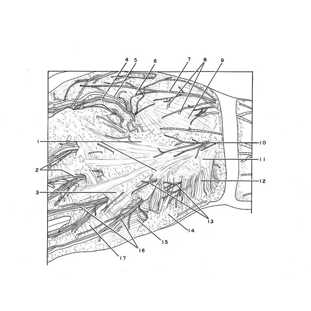
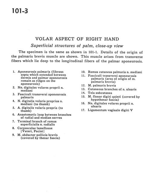
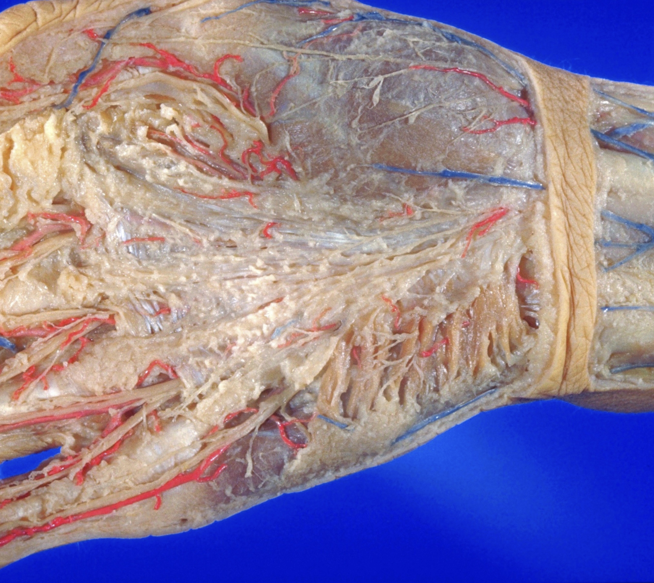
The specimen is the same as shown in 101-1. Details of the origin of the palmaris brevis muscle are shown. This muscle arises from transverse fibres which lie deep to the longitudinal fibres of the palmar aponeurosis.
- Palmar aponeurosis (fibrous septa which extended between dermis and palmar aponeurosis remain as ridges on the aponeurosis)
- Proper palmar digital nerves of median nerve
- Transverse fascicle of palmar aponeurosis
- Proper palmar digital nerve of median nerve (to thumb)
- Proper palmar digital artery (to thumb)
- Anastomotic loop between branches of radial and median nerves
- Terminal branch of superficial branch of radial nerve
- Pacinian corpuscles
- Abductor pollicis brevis muscle (covered by thenar fascia)
- Palmar cutaneous branch of median nerve
- Transverse fascicle of palmar aponeurosis (area of origin of palmaris brevis muscle)
- Palmaris brevis muscle
- Cutaneous branches of of ulnar nerve
- Superficial fascia
- Flexor digiti minimi muscle (covered by hypothenar fascia)
- Proper palmar digital nerves of ulnar nerve
- Ligament of digital sheath V