X CLOSE

Exploration of the brain from its superior aspect

Right centrum semiovale

Stanford holds the copyright to the David L. Bassett anatomical images and has assigned Creative Commons license Attribution-Share Alike 4.0 International to all of the images.
For additional information regarding use and permissions, please contact Dr. Drew Bourn at dbourn@stanford.edu.

Image #10-1
Bassett ImageDiagram Image
Legend Image
Exploration of the brain from its superior aspect
Right centrum semiovale
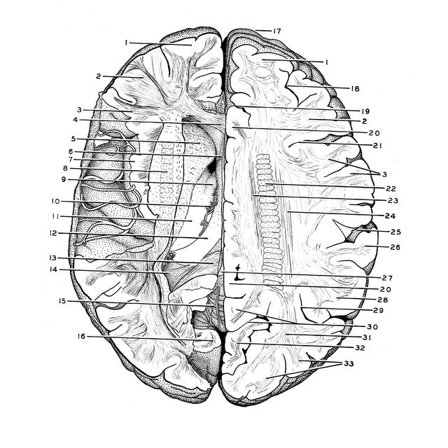
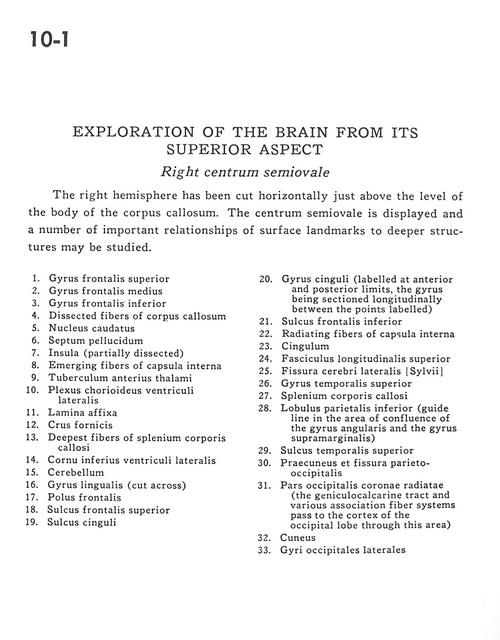
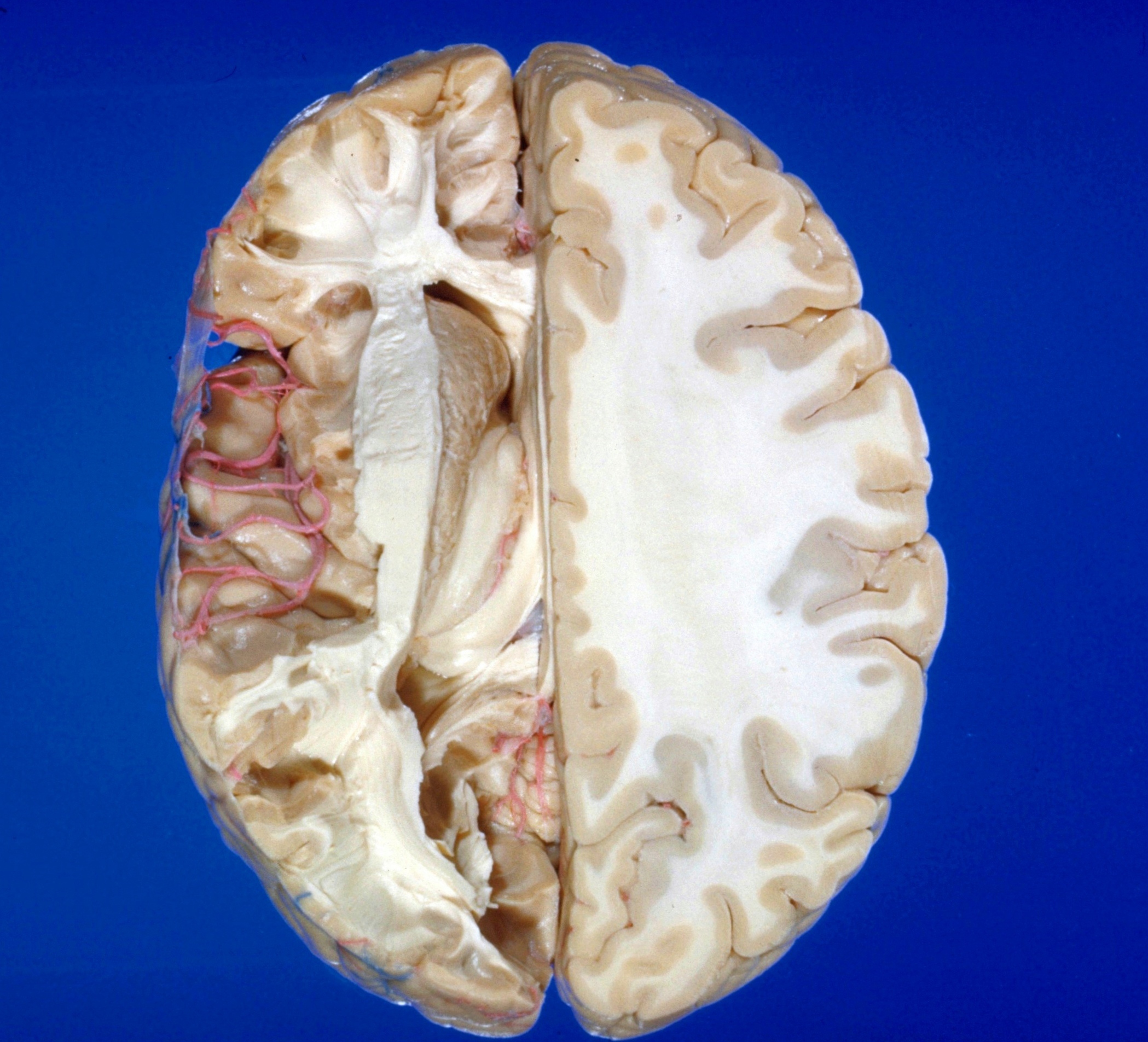
The right hemisphere has been cut horizontally just above the level of the body of the corpus callosum. The centrum semiovale is displayed and a number of important relationships of surface landmarks to deeper structures may be studied.
1 . Superior frontal gyrus
2 . Medial frontal gyrus
3 . Inferior frontal gyrus
4 . Dissected fibers of corpus callosum
5 . Caudate nucleus
6 . Septum pellucidum
7 . Insula (partially dissected)
8 . Emerging fibers of internal capsule
9 . Anterior tubercle of thalamus
10 . Choroid plexus lateral ventricle
11 . Lamina affixa
12 . Fornix (crus)
13 . Deepest fibers of corpus callosum (splenium)
14 . Inferior horn of lateral ventricle
15 . Cerebellum
16 . Lingual gyrus (cut across)
17 . Frontal pole
18 . Superior frontal sulcus
19 . Cingulate sulcus
20 . Cingulate gyrus (labeled at anterior and posterior limits, the gyrus being sectioned longitudinally between the points labeled)
21 . Inferior frontal sulcus
22 . Radiating fibers of internal capsule
23 . Cingulum
24 . Superior longitudinal fasciculus
25 . Lateral cerebral fissure (sylvian)
26 . Superior temporal gyrus
27 . Corpus callosum (splenium)
28 . Inferior parietal lobule (guide line in the area of confluence of the angular gyrus and the supramarginal gyrus)
29 . Superior temporal sulcus
30 . Precuneus and parieto-occipital fissure
31 . Occipital part corona radiata (the geniculocalcarine tract and various association fiber systems pass to the cortex of the occipital lobe through this area)
32 . Cuneus
33 . Lateral occipital gyri