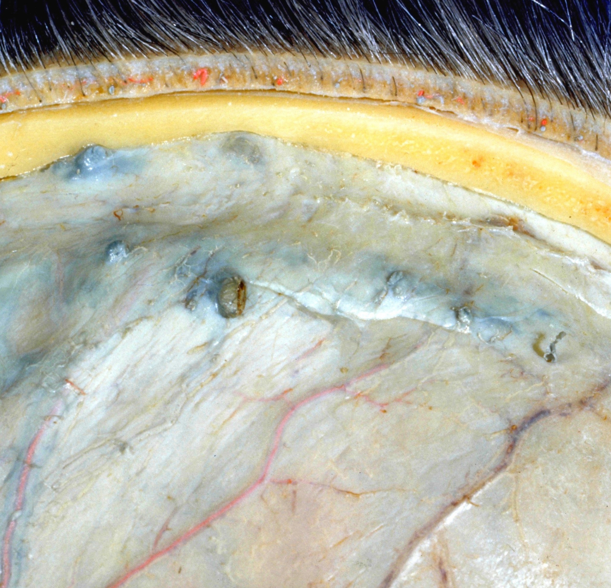
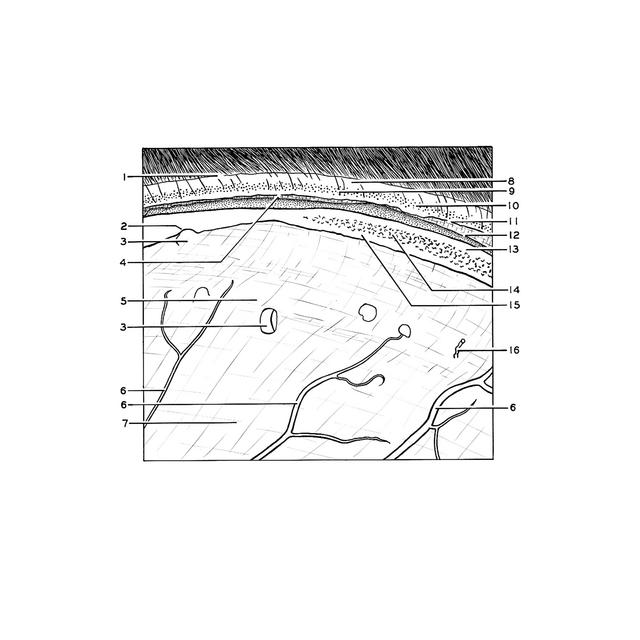
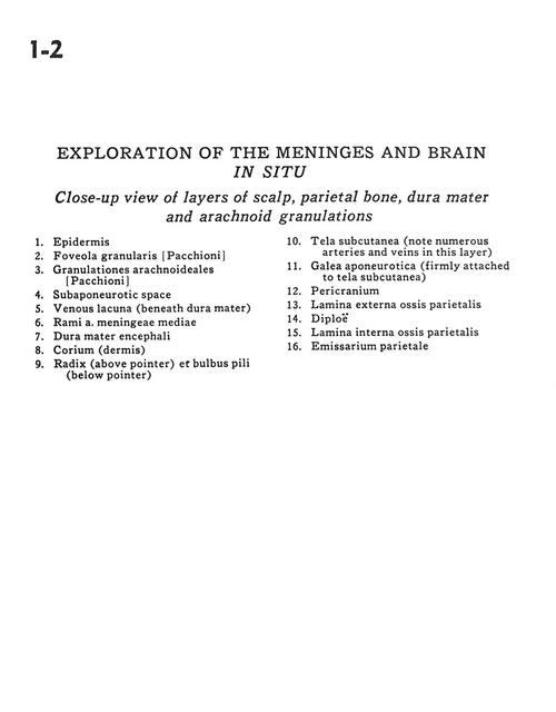
Exploration of the meninges and brain in situ
Close-up view of layers of scalp, parietal bone, dura mater and arachnoid granulations
Stanford holds the copyright to the David L. Bassett anatomical images and has assigned
Creative Commons license Attribution-Share Alike 4.0 International to all of the images.
For additional information regarding use and permissions,
please contact Dr. Drew Bourn at dbourn@stanford.edu.
Image #1-2
 |  | ||||||||||||||||||||||||||||||||||||
 |
|