
Volar aspect of forearm
Nerve supply to flexor digitorum profundus muscle; volar interosseous artery and nerve
Stanford holds the copyright to the David L. Bassett anatomical images and has assigned
Creative Commons license Attribution-Share
Alike 4.0 International to all of the images.
For additional information regarding use and permissions,
please contact Dr. Drew Bourn at dbourn@stanford.edu.
Image #99-1
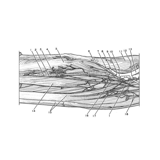
Volar aspect of forearm
Nerve supply to flexor digitorum profundus muscle; volar interosseous artery and nerve
The flexor pollicis longus muscle has been retracted laterally to reveal the muscular branches of the interosseous nerve which supply the medial part of the muscle. Nerves to the flexor digitorum profundus have been exposed, particularly to that part of the muscle which inserts on the index finger.
- Anterior interosseous nerve
- Anterior interosseous artery
- Anastomotic loop between two muscular branches of anterior interosseous nerve
- Radius
- Flexor pollicis longus muscle (retracted laterally)
- Pronator teres muscle (insertion)
- Muscular branch anterior interosseous nerve (to flexor pollicis longus muscle)
- Median artery (large)
- Ulnar artery
- Dorsal interosseous artery
- Supinator muscle
- Muscular branch ulnar artery
- Ulnar artery (proximal part)
- Flexor digitorum profundus muscle (to digit II)
- Flexor digitorum profundus muscle (to digits III-V)
- Muscular branch anterior interosseous nerve (to parts of flexor digitorum profundus muscle)
- Muscular branch anterior interosseous nerve (to parts of flexor digitorum profundus muscle)
- Muscular branch of ulnar nerve (to medial part of flexor digitorum profundus muscle)