X CLOSE

Shoulder

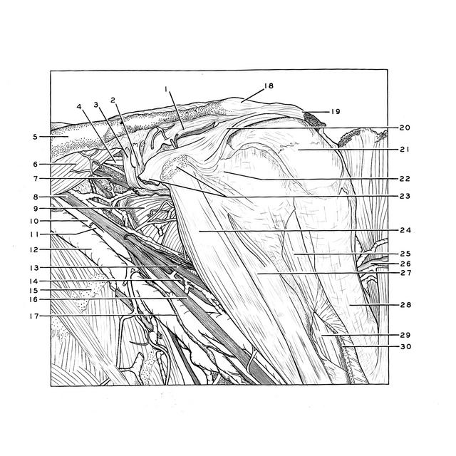
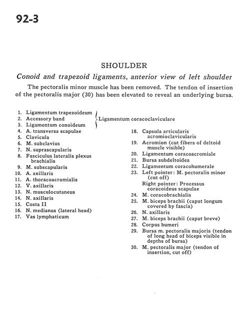
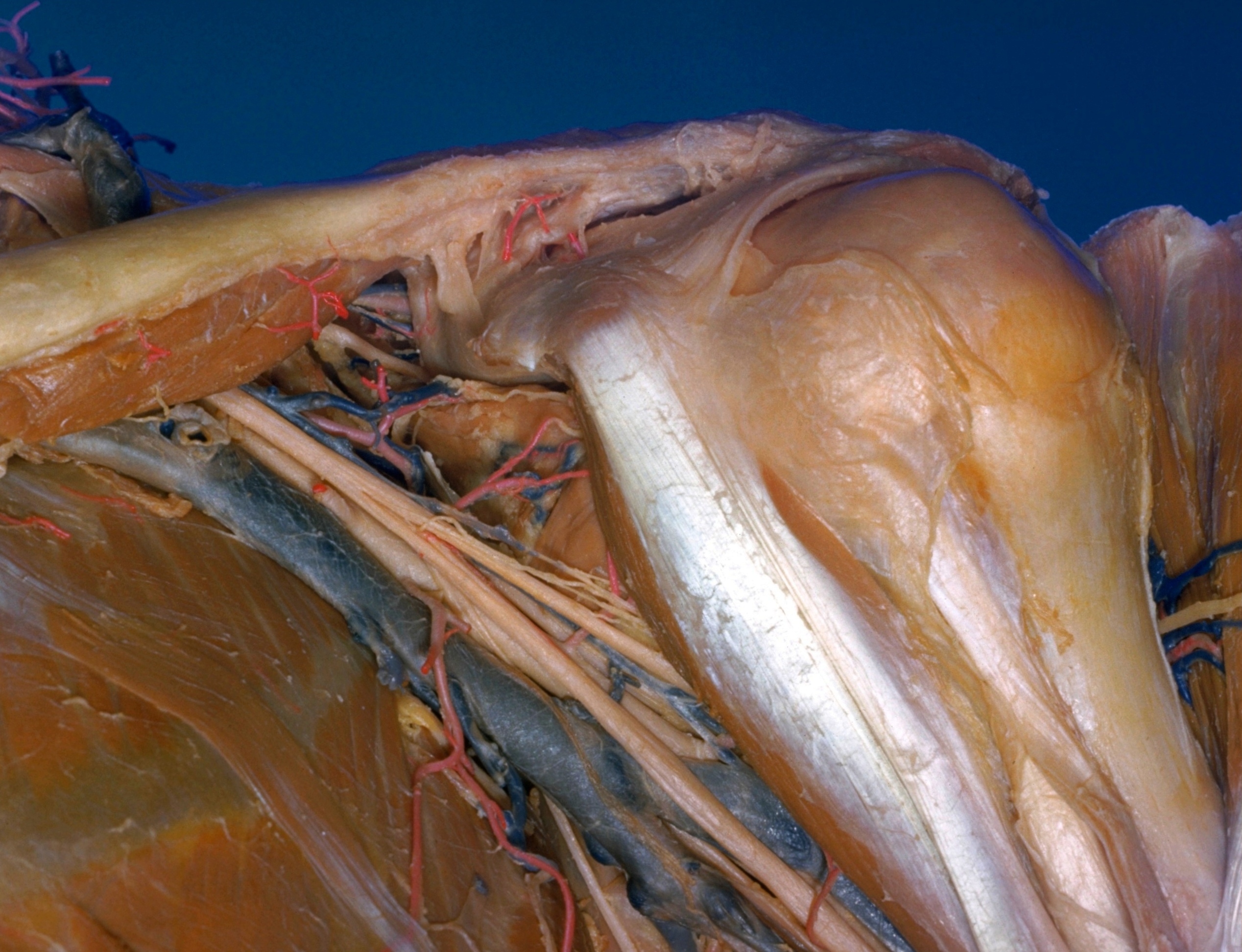
Conoid and trapezoid ligaments, anterior view of left shoulder

Stanford holds the copyright to the David L. Bassett anatomical images and has assigned Creative Commons license Attribution-Share Alike 4.0 International to all of the images.
For additional information regarding use and permissions, please contact Dr. Drew Bourn at dbourn@stanford.edu.

Image #92-3
Bassett ImageDiagram Image
Legend Image
Shoulder
Conoid and trapezoid ligaments, anterior view of left shoulder
The pectoralis minor muscle has been removed. The tendon of insertion of the pectoralis major (30) has been elevated to reveal an underlying bursa.
1 . Trapezoid ligament
2 . Accessory band
3 . Conoid ligament (92-4 make up the coracoclavicular ligament)
4 . Transverse scapular artery
5 . Clavicle
6 . Subclavius muscle
7 . Suprascapular nerve
8 . Lateral cord of brachial plexus
9 . Subscapularis muscle
10 . Axillary artery
11 . Thoracoacromial artery
12 . Axillary vein
13 . Musculocutaneous nerve
14 . Axillary nerve
15 . Rib II
16 . Median nerve (lateral head)
17 . Lymph vessel
18 . Acromioclavicular joint capsule
19 . Acromion (cut fibers of deltoid muscle visible)
20 . Coracoacromial ligament
21 . Subdeltoid bursa
22 . Coracohumeral ligament
23 . Left pointer: Pectoralis minor muscle (cut off) Right pointer: Coracoid process of scapula
24 . Coracobrachialis muscle
25 . Biceps brachii muscle (long head covered by fascia)
26 . Axillary nerve
27 . Biceps brachii muscle (short head)
28 . Body of humerus
29 . Bursa pectoralis major muscle (tendon of long head of biceps visible in depths of bursa)
30 . Pectoralis major muscle (tendon of insertion, cut off)