X CLOSE

Shoulder

Left subdeltoid bursa

Stanford holds the copyright to the David L. Bassett anatomical images and has assigned Creative Commons license Attribution-Share Alike 4.0 International to all of the images.
For additional information regarding use and permissions, please contact Dr. Drew Bourn at dbourn@stanford.edu.

Image #92-2
Bassett ImageDiagram Image
Legend Image
Shoulder
Left subdeltoid bursa
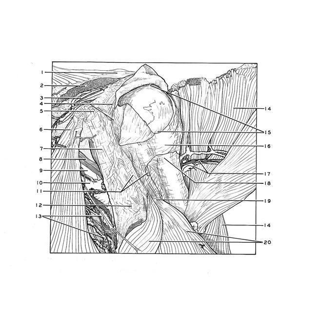
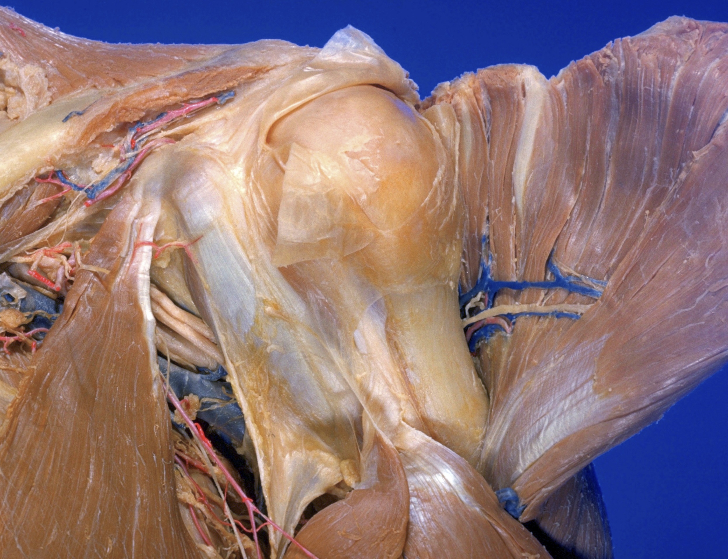
The left deltoid muscle has been cut from its acromial and clavicular origins and reflected laterally. The subdeltoid bursa (15) has been opened. Continuity of the pectoral fascia with the fascia over the coracobrachialis muscle is visible at 12. A thin layer of fascia has been removed from the deep surface of the deltoid muscle to expose the axillary nerve (17) and vessels.
1 . Position of acromioclavicular joint
2 . Deltoid muscle (divided at clavicular origin)
3 . Acromial branch thoracoacromial artery (note articular branch of lateral anterior thoracic nerve accompanying this vessel)
4 . Upper pointer: Coracoacromial ligament Lower pointer: Coracohumeral ligament
5 . Coracoid process of scapula
6 . Pectoralis minor muscle
7 . Coracobrachialis muscle
8 . Musculocutaneous nerve
9 . Axillary artery
10 . Axillary vein
11 . Left pointer: Biceps brachii muscle (short head covered by fascia) Right pointer: Biceps brachii muscle (tendon of long head visible beneath fascia)
12 . Area of confluence of pectoral fascia with fascia of coracobrachialis muscle
13 . Axillary lymph nodes (central group)
14 . Deltoid muscle (reflected laterally)
15 . Subdeltoid bursa (this bursa extends beneath acromial process superiorly and thus forms a common subdeltoid-subacromial bursa)
16 . Fascia covering joint capsule of humerus
17 . Left pointer: Posterior circumflex artery of humerus Right pointer: Axillary nerve
18 . Surgical neck of humerus
19 . Tendon of insertion of pectoralis major muscle
20 . Pectoralis major muscle (reflected laterally and inferiorly)