Exploration of the brain from its superior aspect
Close-up view; relations of posterior horn of lateral ventricle
Stanford holds the copyright to the David L. Bassett anatomical images and has assigned
Creative Commons license Attribution-Share
Alike 4.0 International to all of the images.
For additional information regarding use and permissions,
please contact Dr. Drew Bourn at dbourn@stanford.edu.
Image #9-5
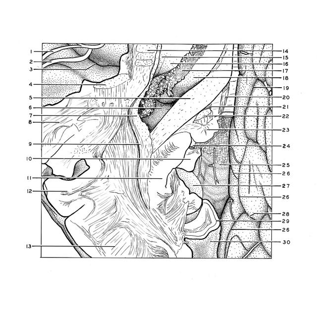
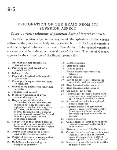
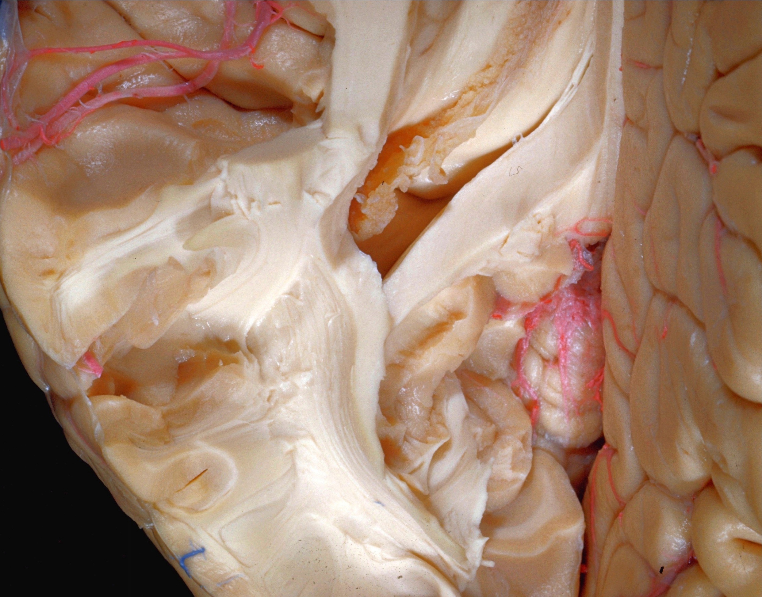
Exploration of the brain from its superior aspect
Close-up view; relations of posterior horn of lateral ventricle
Detailed relationships in the region of the splenium of the corpus callosum, the junction of the body and posterior horn of the lateral ventricle and the occipital lobe are illustrated. Boundaries of the opened ventricle are clearly visible in the upper central part of the view. The line of Gennari appears in the cut section of the lingual gyrus (29).
- Anterior parietal branch of middle cerebral artery
- Posterior parietal branch of middle cerebral artery
- Sulcus circularis
- Superior longitudinal fasciculus (cut across)
- Cut edge of corpus callosum lateral to splenium
- Bulbus posterior horn of lateral ventricle
- Tapetum (cut across)
- Medullary substance of superior temporal gyrus
- Occipitothalamic radiations (Gratiolet) (Note: This stratum includes not only the geniculocalcarine tract but also a system of fibers, the inferior occipitofrontal fasciculus, which can be traced forward toward the frontal lobe in a path lateral to the lentiform nucleus)
- Cut section of the forward continuation of the lingual gyrus which becomes continuous in this region with the hippocampal gyrus
- Gray matter lying in the deepest part of the calcarine fissure
- Dissected portion of angular gyrus of inferior parietal lobule
- Medullary substance of superior occipital gyrus
- Internal capsule
- Stria terminalis
- Lamina affixa
- Choroid plexus lateral ventricle
- Fornix (crus)
- Subparietal sulcus (continuous anteriorly with cingulate sulcus)
- Medial longitudinal stria
- Lateral longitudinal stria
- Cingulum (cut across)
- Limbic lobe (downward continuation around splenium of cingulate gyrus previously seen)
- Posterior cerebral artery in depths of calcarine fissure
- Superior surface of cerebellum facing transverse cerebral fissure (meninges and branches of superior cerebellar artery are visible on surface)
- Calcarine fissure
- Convolution of lower part of lingual gyrus exposed in depths of a minor sulcus
- Parieto-occipital fissure right
- Cut section of visual cortex in lingual gyrus (note line of Gennari, hence the name striate cortex often applied to the visual cortex)
- Surface of lingual gyrus in depths of calcarine fissure