X CLOSE

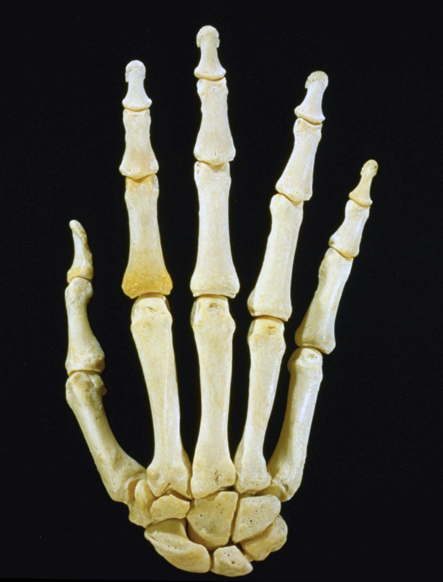
Osteology

Articulated bones of right hand, dorsal view

Stanford holds the copyright to the David L. Bassett anatomical images and has assigned Creative Commons license Attribution-Share Alike 4.0 International to all of the images.
For additional information regarding use and permissions, please contact Dr. Drew Bourn at dbourn@stanford.edu.

Image #87-3
Bassett ImageDiagram Image
Legend Image
Osteology
Articulated bones of right hand, dorsal view
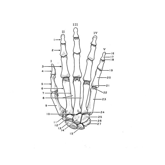
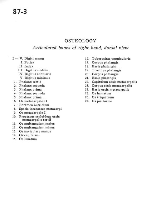
I-V. Digits of the hand I. Thumb H. Index III. Long IV. Ring V. Little.
I-V . Digits of the hand
I . Thumb
II . Index
III . Long
IV . Ring
V . Little
1 . Distal phalanx
2 . Middle phalanx
3 . Proximal phalanx
4 . Middle phalanx
5 . Proximal phalanx
6 . Metacarpal II
7 . Nutritive foramen
8 . Interosseous metacarpal space
9 . Metacarpal I
10 . Styloid process of third metacarpal
11 . Trapezium bone
12 . Trapezoid bone
13 . Scaphoid bone
14 . Capitate bone
15 . Lunate bone
16 . Tuberosity of distal phalanx
17 . Body of phalanx
18 . Base of phalanx
19 . Joint of phalanx
20 . Body of phalanx
21 . Base of phalanx
22 . Head of metacarpal
23 . Body of metacarpal
24 . Base of metacarpal
25 . Hamate bone
26 . Triquetral bone
27 . Pisiform bone