Dissection of head and neck from a posterior approach
Pharyngeal plexus of nerves; ascending pharyngeal artery
Stanford holds the copyright to the David L. Bassett anatomical images and has assigned
Creative Commons license Attribution-Share
Alike 4.0 International to all of the images.
For additional information regarding use and permissions,
please contact Dr. Drew Bourn at dbourn@stanford.edu.
Image #82-3
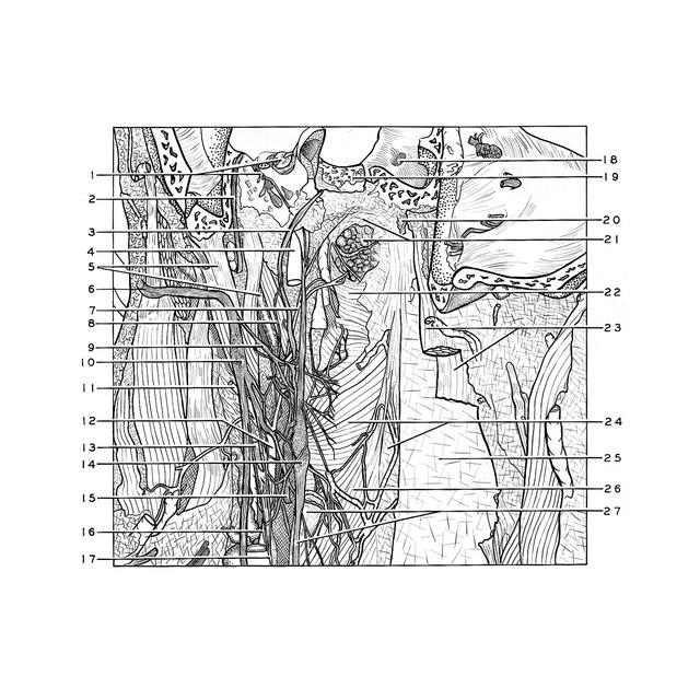
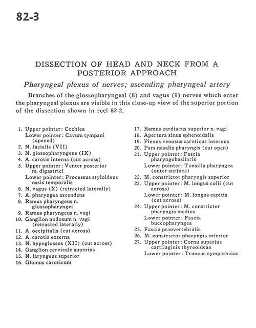
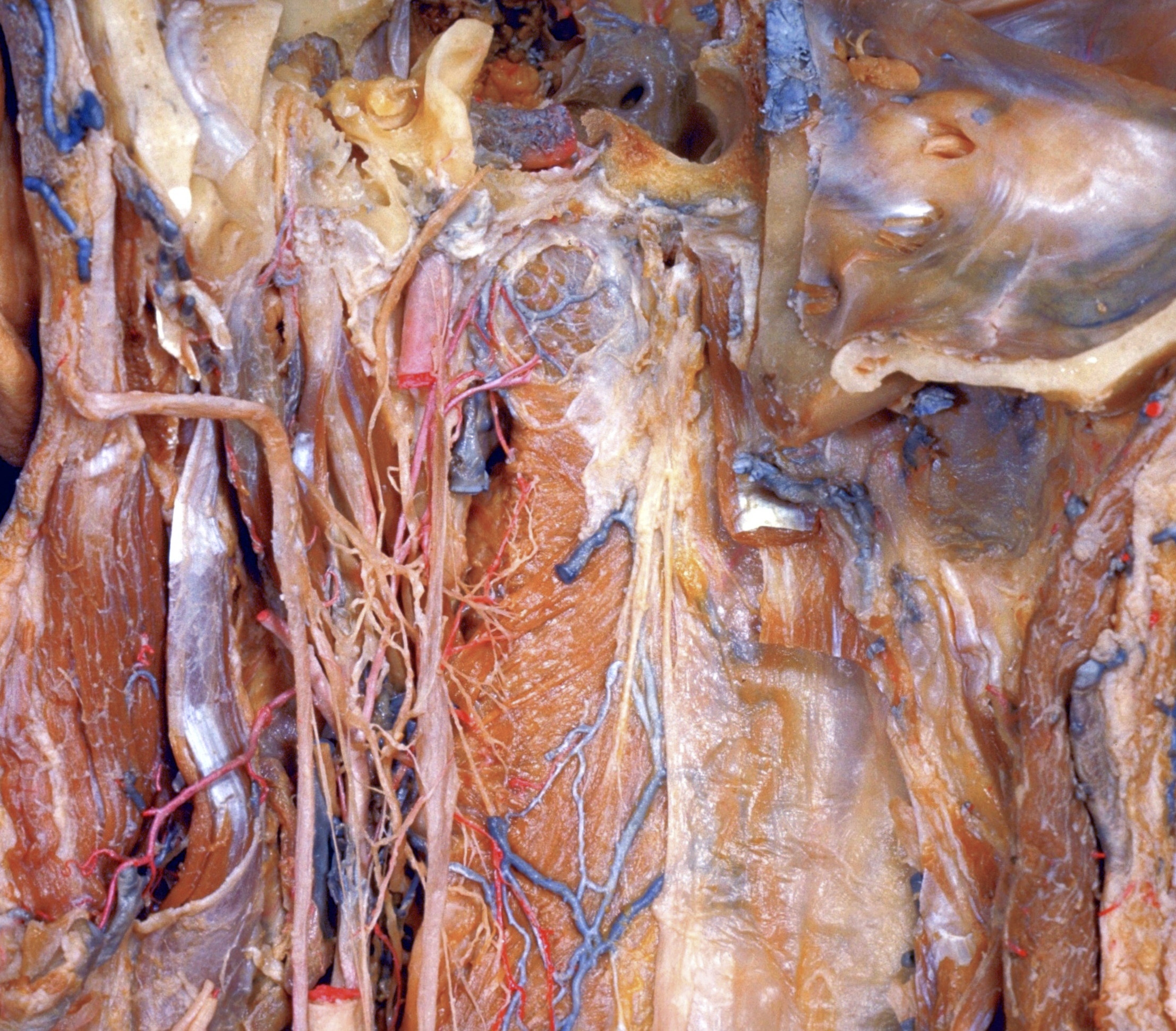
Dissection of head and neck from a posterior approach
Pharyngeal plexus of nerves; ascending pharyngeal artery
Branches of the glossopharyngeal (8) and vagus (9) nerves which enter the pharyngeal plexus are visible in this close-up view of the superior portion of the dissection shown in reel 82-2.
- Upper pointer: Cochlea Lower pointer: Tympanic cavity (opened)
- Facial nerve (VII)
- Glossopharyngeal nerve (IX)
- Internal carotid artery (cut across)
- Upper pointer: Posterior belly of digastric muscle Lower pointer: Styloid process temporal bone
- Vagus nerve (X) (retracted Iaterally)
- Ascending pharyngeal artery
- Pharyngeal branch glossopharyngeal nerve
- Pharyngeal branch vagus nerve
- Nodose ganglion of vagus nerve (retracted laterally)
- Occipital artery (cut across)
- External carotid artery
- Hypoglossal nerve (XII) (cut across)
- Superior cervical ganglion
- Superior laryngeal nerve
- Carotid body
- Superior cardiac branch vagus nerve
- Aperture of sphenoid sinus
- Internal carotid venous plexus
- Nasal part pharynx (cut open)
- Upper pointer: Pharyngobasilar fascia Lower pointer: Pharyngeal tonsil (outer surface)
- Superior pharyngeal constrictor muscle
- Upper pointer: Longus colli muscle (cut across) Lower pointer: Longus capitis muscle (cut across)
- Upper pointer: Middle pharyngeal constrictor muscle Lower pointer: Buccopharyngeal fascia
- Prevertebral fascia
- Inferior pharyngeal constrictor muscle
- Upper pointer: Superior horn thyroid cartilage Lower pointer: Sympathetic trunk