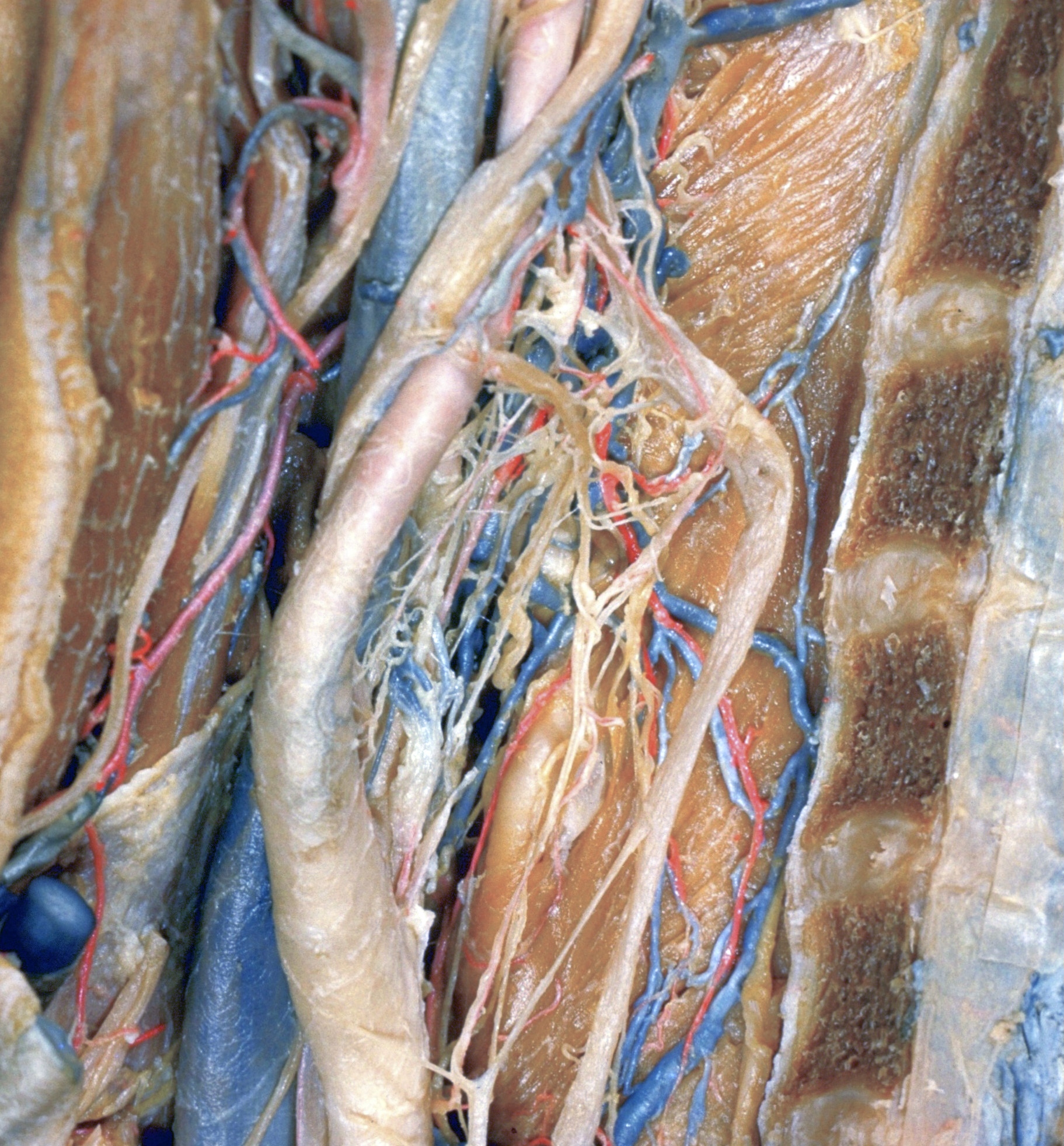
Dissection of head and neck from a posterior approach
Blood vessels and nerves to left carotid body
Stanford holds the copyright to the David L. Bassett anatomical images and has assigned
Creative Commons license Attribution-Share
Alike 4.0 International to all of the images.
For additional information regarding use and permissions,
please contact Dr. Drew Bourn at dbourn@stanford.edu.
Image #82-1
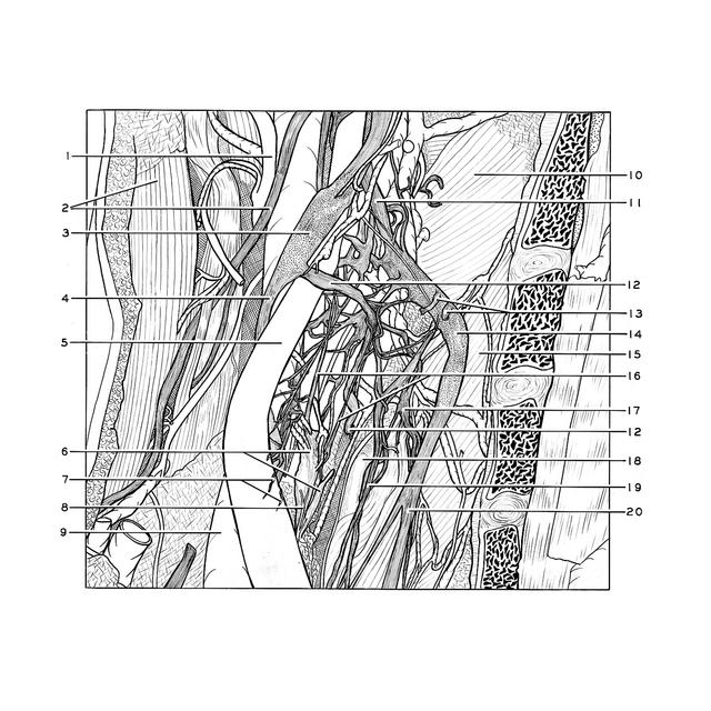
Dissection of head and neck from a posterior approach
Blood vessels and nerves to left carotid body
The internal carotid artery and vagus nerve have been retracted posterolaterally and the superior cervical ganglion retracted posteromedially.
- Occipital artery
- Upper pointer: Sternocleidomastoid muscle Lower pointer: Accessory nerve (XI)
- Nodose ganglion of vagus nerve
- Vagus nerve (X) (retracted posterolaterally)
- Internal carotid artery (retracted posterolaterally)
- Upper pointer: Carotid body Lower pointer: Carotid body (accessory)
- Nerve filaments entering wall of Internal carotid artery at site of carotid sinus
- Artery which supplies carotid body
- Internal jugular vein
- Middle pharyngeal constrictor muscle
- Internal carotid nerve
- Superior laryngeal nerve
- Gray rami communicantes (to cervical nerve I-II)
- Superior cervical ganglion
- Inferior pharyngeal constrictor muscle
- Nerves to carotid body (branches of glossopharyngeal nerve (IX))
- Pharyngeal plexus vagus nerve
- Superior horn thyroid cartilage
- Superior cardiac branch vagus nerve
- Sympathetic trunk