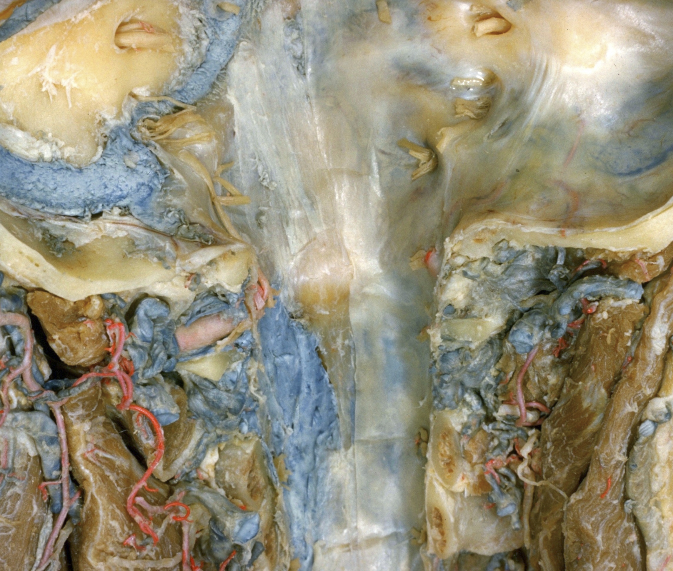
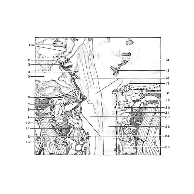
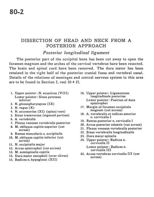
Dissection of head and neck from a posterior approach
Posterior longitudinal ligament
Stanford holds the copyright to the David L. Bassett anatomical images and has assigned
Creative Commons license Attribution-Share
Alike 4.0 International to all of the images.
For additional information regarding use and permissions,
please contact Dr. Drew Bourn at dbourn@stanford.edu.
Image #80-2
Dissection of head and neck from a posterior approach
Posterior longitudinal ligament
The posterior part of the occipital bone has been cut away to open the foramen magnum and the arches of the cervical vertebrae have been resected. The brain and spinal cord have been removed. The dura mater has been retained in the right half of the posterior cranial fossa and vertebral canal. Details of the relations of meninges and central nervous system in this area are to be found in Section I, reel 32-4 ff.
- Upper pointer: Vestibulocochlear nerve (VIII) Lower pointer: Inferior petrosal sinus
- Glossopharyngeal nerve (IX)
- Vagus. nerve (X)
- Accessory nerve (XI) (spinal root)
- Transverse sinus (sigmoid portion)
- Vertebral artery
- Posterior vertebral venous plexus
- Superior oblique capitis muscle (cut across)
- Muscular branch occipital artery
- Inferior oblique capitis muscle (cut across)
- Greater occipital nerve
- Arch of axis (cut across)
- Semispinalis capitis muscle
- Dura mater (over clivus)
- Roots of hypoglossal nerve (XII)
- Upper pointer: Posterior longitudinal ligament Lower pointer: Position of dens (axis)
- Margin of foramen magnum (cut across)
- Vertebral artery and ventral roots of cervical nerve I
- Posterior branch cervical nerve I
- Posterior arch of atlas (cut across)
- Posterior vertebral venous plexus
- Longitudinal vertebral sinus
- Dura mater
- Upper pointer: Roots cervical nerve II Lower pointer: Roots cervical nerve III
- Arch of cervical vertebra III (cut across)