Dissection of head and neck from a posterior approach
Superficial structures; fascia colli
Stanford holds the copyright to the David L. Bassett anatomical images and has assigned
Creative Commons license Attribution-Share
Alike 4.0 International to all of the images.
For additional information regarding use and permissions,
please contact Dr. Drew Bourn at dbourn@stanford.edu.
Image #78-5
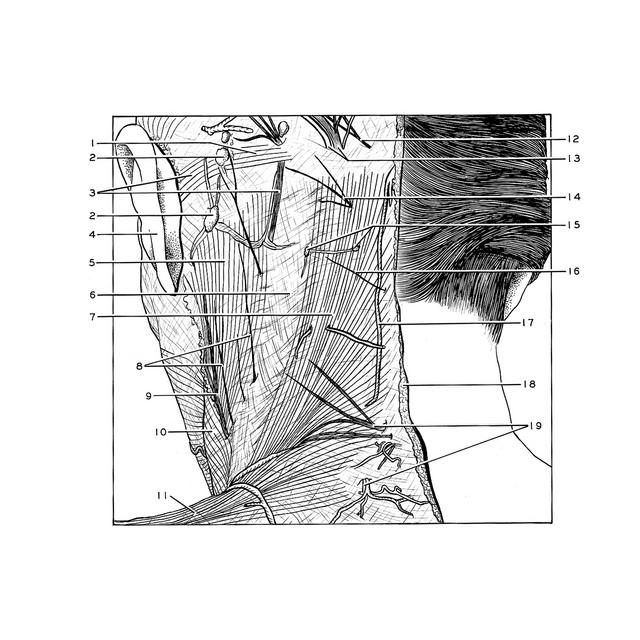
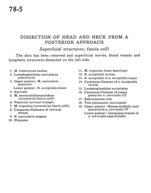
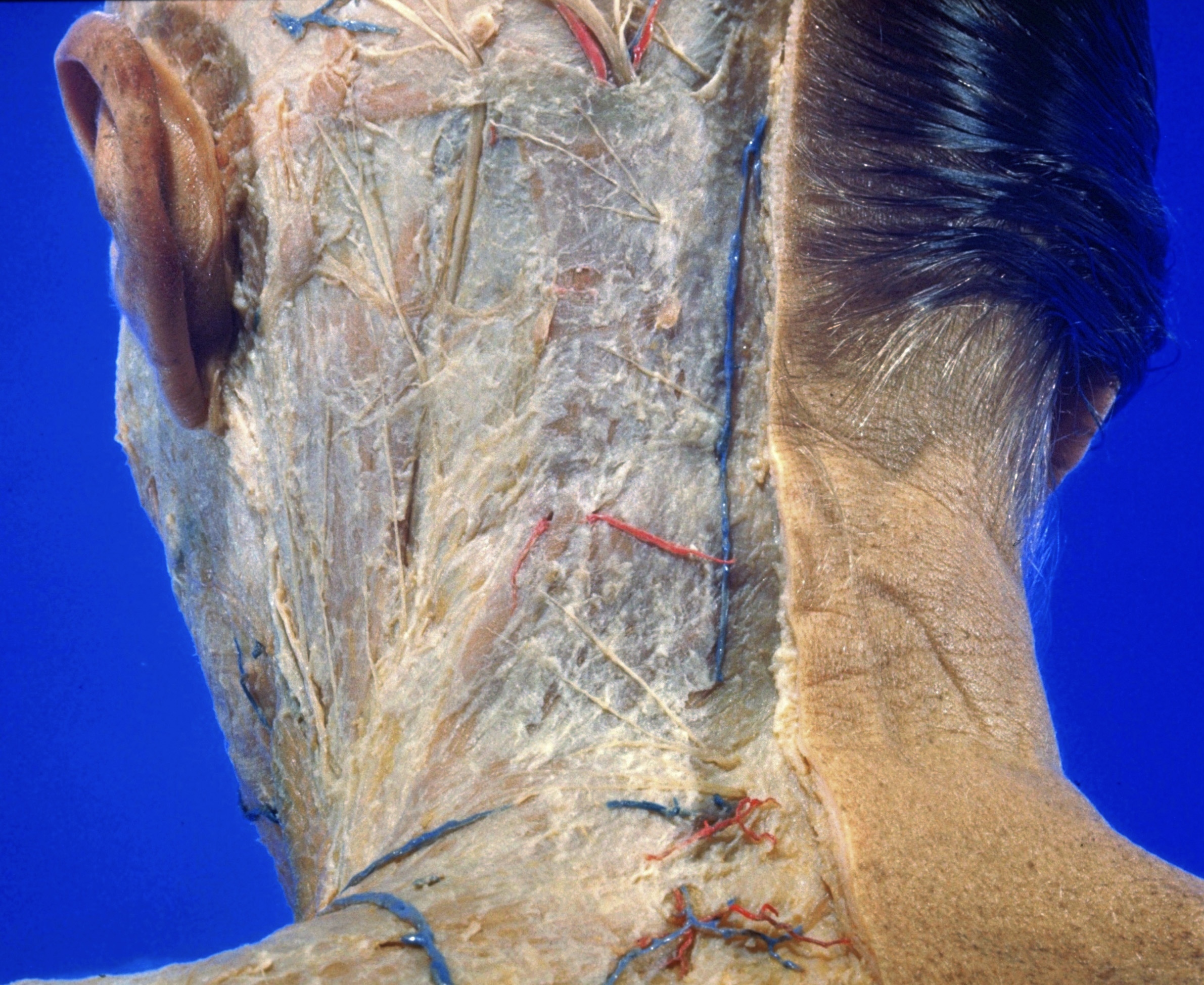
Dissection of head and neck from a posterior approach
Superficial structures; fascia colli
The skin has been removed and superficial nerves, blood vessels and lymphatic structures dissected on the left side.
- Transverse nuchal muscle
- Posterior auricular lymph nodes
- Upper pointer: Posterior auricular muscle Lower pointer: Lesser occipital nerve
- Auricle
- Sternocleidomastoid muscle (covered by superficial fascia)
- Posterior cervical triangle
- Trapezius muscle (covered by superficial fascia)
- Cutaneous filaments of cervical plexus
- Greater auricular nerve
- Platysma
- Trapezius muscle (near insertion)
- Third occipital nerve
- Occipital artery and major occipital nerve
- Cutaneous filament of third occipital nerve
- Occipital lymph nodes
- Cutaneous filament of posterior branch cervical nerve III
- Subcutaneous vein
- Superficial fascia (sectioned)
- Upper pointer: Medial branches of posterior cervical nerve IV Lower pointer: Cutaneous branch of superficial cervical artery