X CLOSE

Dissection of anterior and lateral regions of neck

Larynx; thyroarytenoid and lateral cricoarytenoid muscles; anastomosis of superior and inferior laryngeal nerves, left anterolateral view

Stanford holds the copyright to the David L. Bassett anatomical images and has assigned Creative Commons license Attribution-Share Alike 4.0 International to all of the images.
For additional information regarding use and permissions, please contact Dr. Drew Bourn at dbourn@stanford.edu.

Image #77-4
Bassett ImageDiagram Image
Legend Image
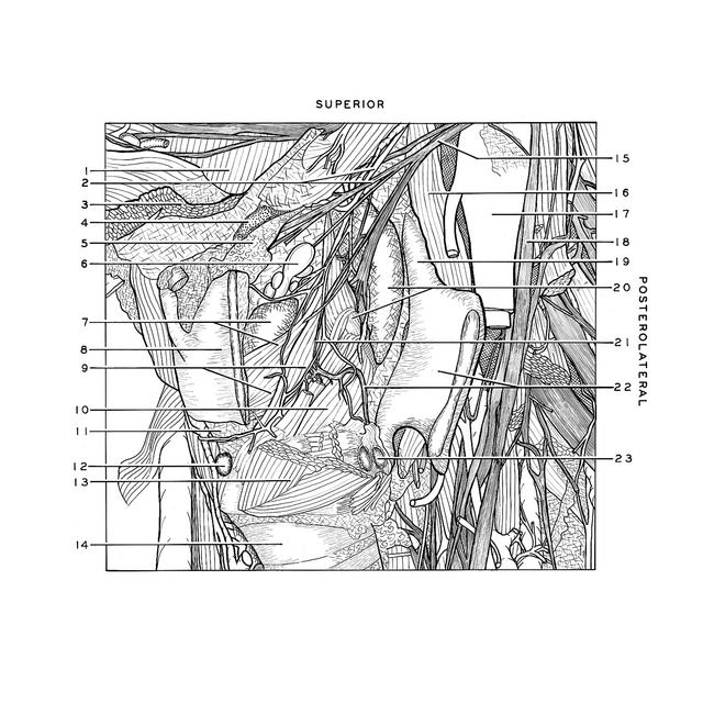
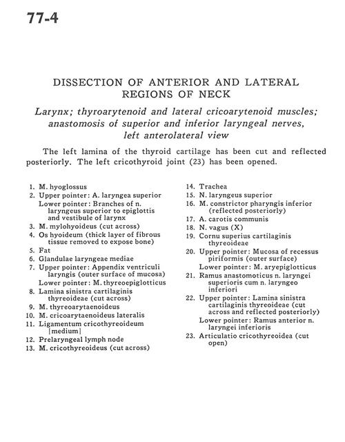
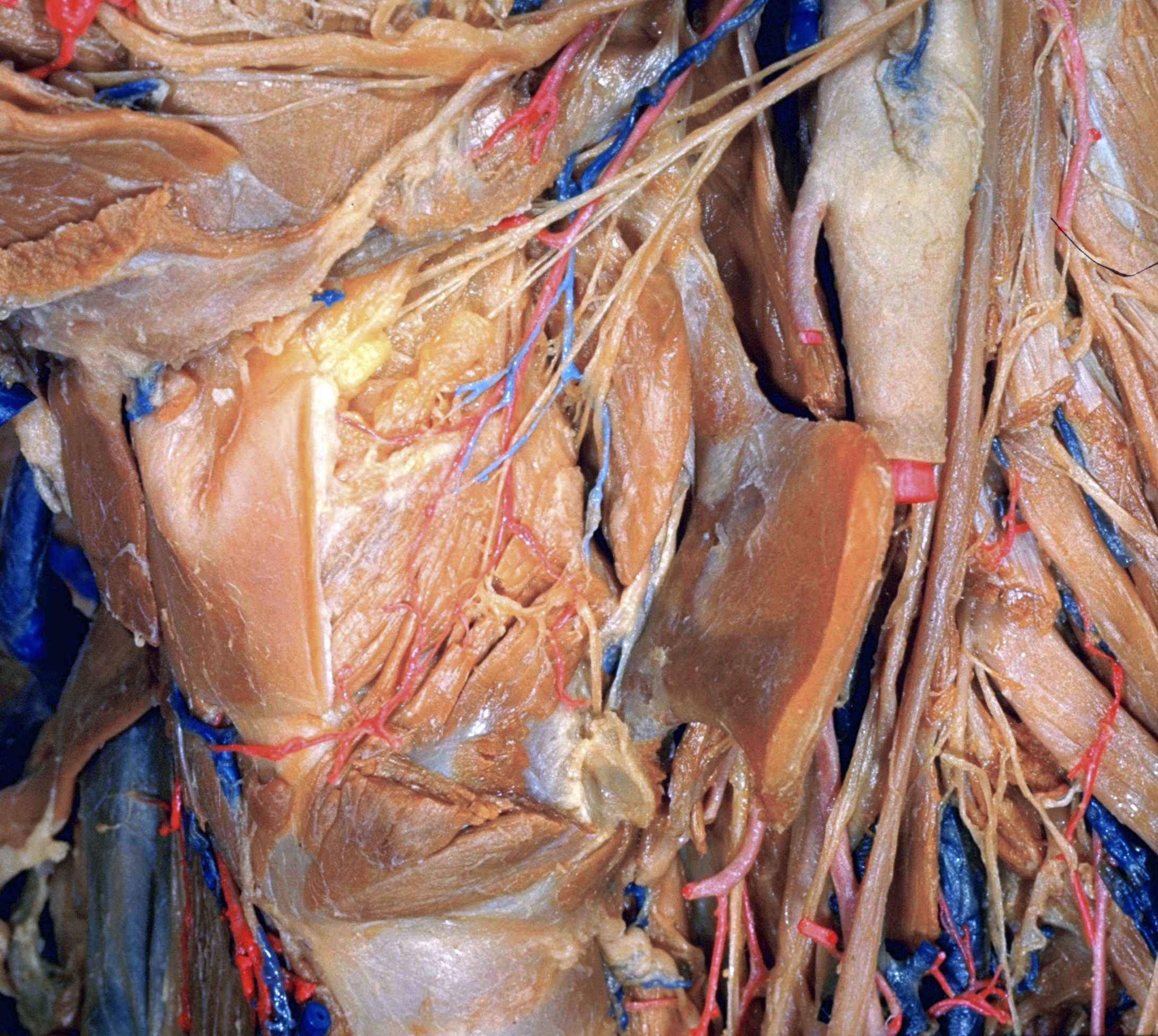
Dissection of anterior and lateral regions of neck
Larynx; thyroarytenoid and lateral cricoarytenoid muscles; anastomosis of superior and inferior laryngeal nerves, left anterolateral view
The left lamina of the thyroid cartilage has been cut and reflected posteriorly. The left cricothyroid joint (23) has been opened.
1 . Hyoglossus muscle
2 . Upper pointer: Superior laryngeal artery Lower pointer: Branches of superior laryngeal nerve to epiglottis and vestibule of larynx
3 . Mylohyoid muscle (cut across)
4 . Hyoid bone (thick layer of fibrous tissue removed to expose bone)
5 . Fat
6 . Middle laryngeal gland
7 . Upper pointer: Appendix laryngeal ventricle (outer surface of mucosa) Lower pointer: Thyroepiglotticus muscle
8 . Left lamina thyroid cartilage (cut across)
9 . Thyroarytenoid muscle
10 . Lateral cricoarytenoid muscle
11 . Cricoarytenoid ligament
12 . Prelaryngeal lymph node
13 . Cricothyroid muscle (cut across)
14 . Trachea
15 . Superior laryngeal nerve
16 . Inferior pharyngeal constrictor muscle (reflected posteriorly)
17 . Common carotid artery
18 . Vagus nerve (X)
19 . Superior horn thyroid cartilage
20 . Upper pointer: Mucosa of piriform recess (outer surface) Lower pointer: Aryepiglotticus muscle
21 . Anastomotic branch superior laryngeal nerve with inferior laryngeal nerve
22 . Upper pointer: Left lamina thyroid cartilage (cut across and reflected posteriorly) Lower pointer: Anterior branch inferior laryngeal nerve
23 . Cricothyroid articulation (cut open)