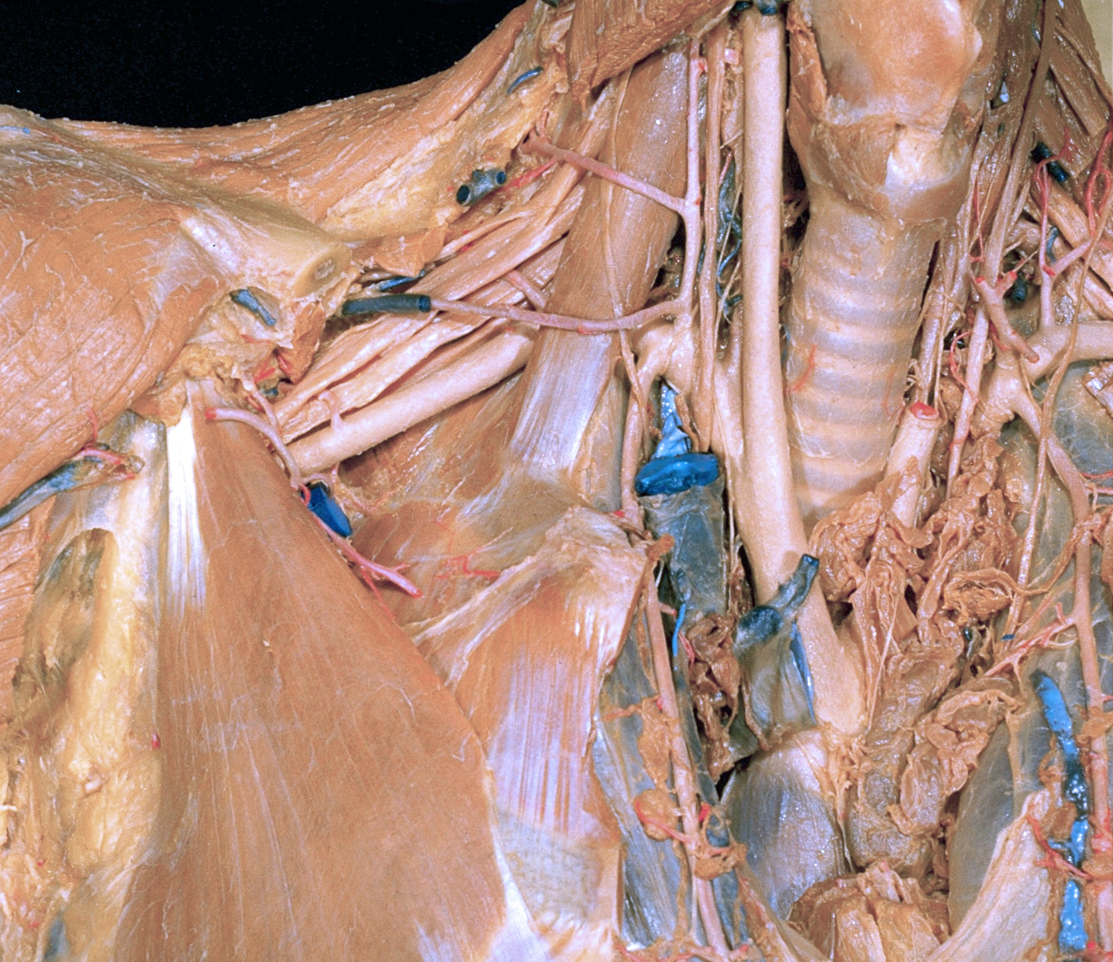
Dissection of anterior and lateral regions of neck
Right phrenic nerve; innominate artery and branches, anterior view
Stanford holds the copyright to the David L. Bassett anatomical images and has assigned
Creative Commons license Attribution-Share
Alike 4.0 International to all of the images.
For additional information regarding use and permissions,
please contact Dr. Drew Bourn at dbourn@stanford.edu.
Image #76-7
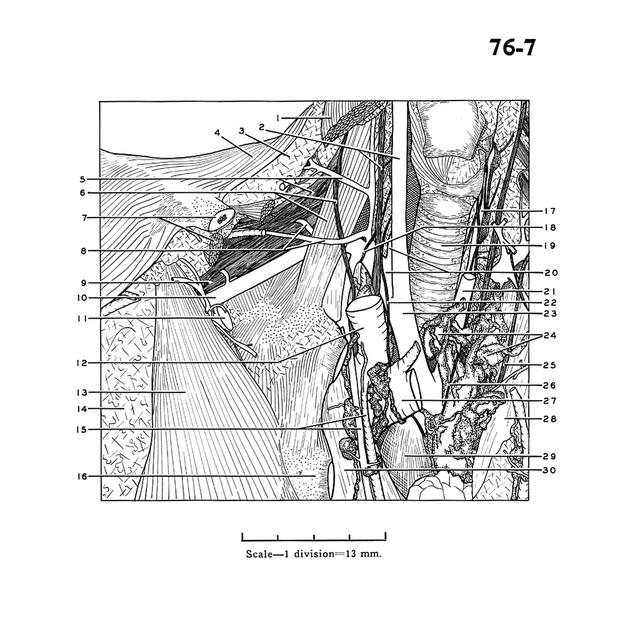
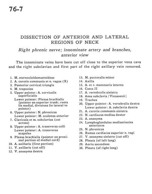
Dissection of anterior and lateral regions of neck
Right phrenic nerve; innominate artery and branches, anterior view
The innominate veins have been cut off close to the superior vena cava and the right subclavian and first part of the right axillary vein removed.
- Sternocleidomastoid muscle
- Common carotid artery and vagus nerve (X)
- Posterior cervical triangle
- Trapezius muscle
- Upper pointer: Superficial cervical artery Lower pointer: Brachial plexus (pointer on superior trunk; roots lie medial, divisions lie lateral to pointer)
- Upper pointer: Phrenic nerve Lower pointer: Anterior scalene muscle
- Clavicle and subclavius muscle (cut across)
- Upper pointer: Transversa colli artery Lower pointer: Transverse scapular artery
- Brachial plexus (pointer on proximal portion of medial cord)
- Axillary artery (first portion)
- Axillary vein (cut off)
- Brachiocephalic vein right
- Pectoralis minor muscle
- Axilla
- Internal thoracic artery and vein
- Rib II
- Vertebral artery left
- Ansa subclavia
- Trachea
- Upper pointer: Vertebral artery right Lower pointer: Subclavian artery right
- Common carotid artery left
- Middle right cardiac nerve
- Brachiocephalic artery
- Anterior mediastinal lymph node
- Phrenic nerve
- Superior cardiac branch of vagus nerve
- Brachiocephalic vein left (cut off)
- Pleura (of left lung)
- Ascending aorta
- Pleura (of right lung)