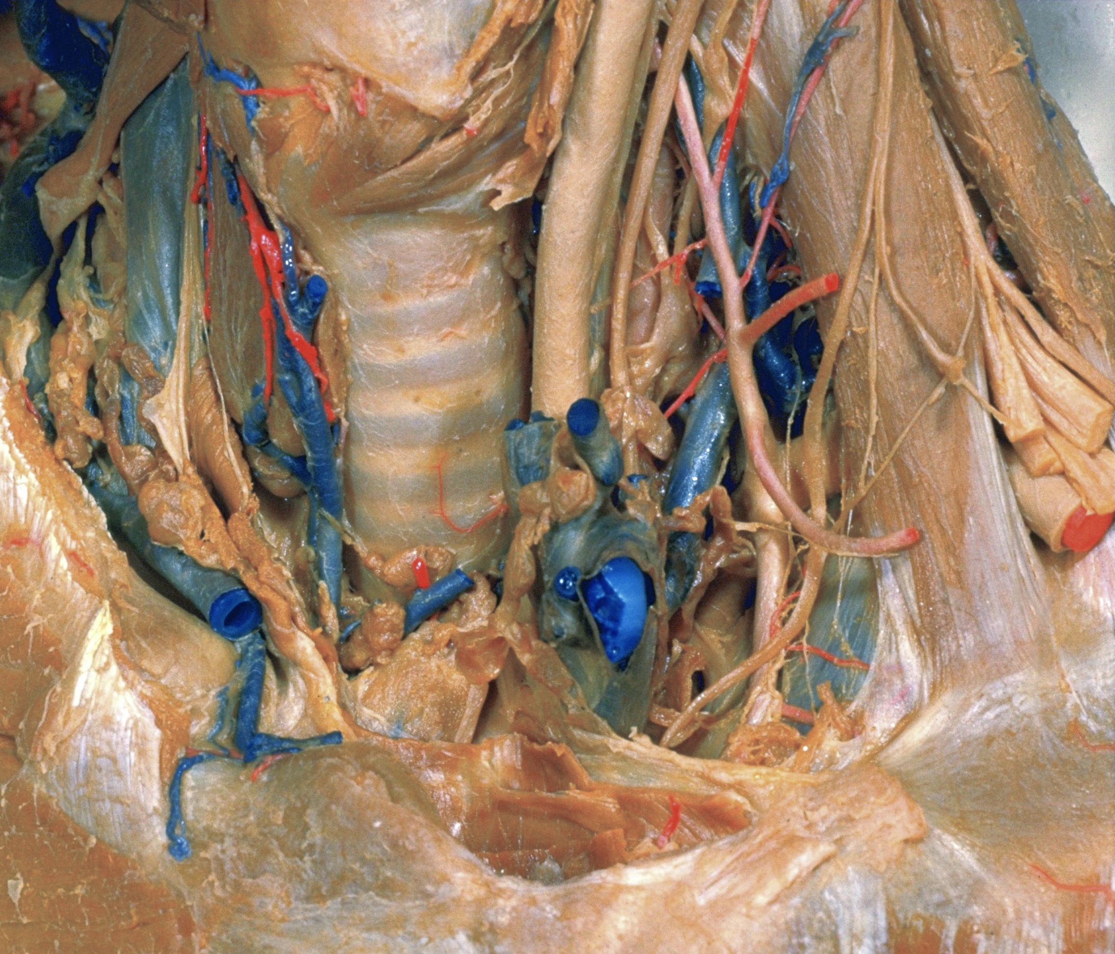
Dissection of anterior and lateral regions of neck
Thoracic duct; structures related to cupula of pleura, left anterolateral view
Stanford holds the copyright to the David L. Bassett anatomical images and has assigned
Creative Commons license Attribution-Share
Alike 4.0 International to all of the images.
For additional information regarding use and permissions,
please contact Dr. Drew Bourn at dbourn@stanford.edu.
Image #76-3
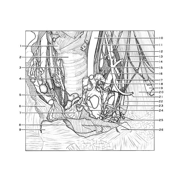
Dissection of anterior and lateral regions of neck
Thoracic duct; structures related to cupula of pleura, left anterolateral view
The internal jugular vein has been cut off and the left innominate vein retracted anteromedially.
- Cricoid cartilage
- Trachea
- Isthmus of thyroid gland (cut across)
- Inferior thyroid vein right
- Sternothyroid muscle
- Sternal extremity of clavicle (covered by joint capsule)
- Thymus
- Articular surface of manubrium of sternum for left clavicle
- Manubrium of sternum
- Common carotid artery
- Vagus nerve (X)
- Ascending cervical artery
- Phrenic nerve
- Superficial cervical artery
- Inferior cervical ganglion
- Vertebral vein
- Thyrocervical trunk
- Thoracic duct
- Subclavian artery
- Anterior scalene muscle
- Transverse scapular artery
- Cupula pleurae
- Internal thoracic (mammary) artery (note internal mammary nerve plexus)
- Anterior and posterior bronchomediastinal lymphatic trunks
- Left internal mammary lymphatic vessels
- Upper pointer: Anonymous vein left (retracted anteromedially) Lower pointer: Costoclavicular ligament