Dissection of anterior and lateral regions of neck
Fascial relations at apex of parietal pleura, left lateral view
Stanford holds the copyright to the David L. Bassett anatomical images and has assigned
Creative Commons license Attribution-Share
Alike 4.0 International to all of the images.
For additional information regarding use and permissions,
please contact Dr. Drew Bourn at dbourn@stanford.edu.
Image #76-2
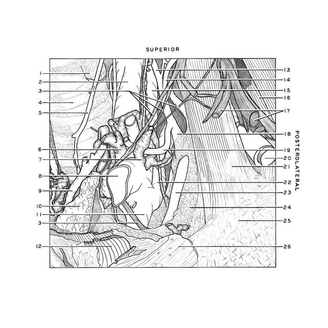
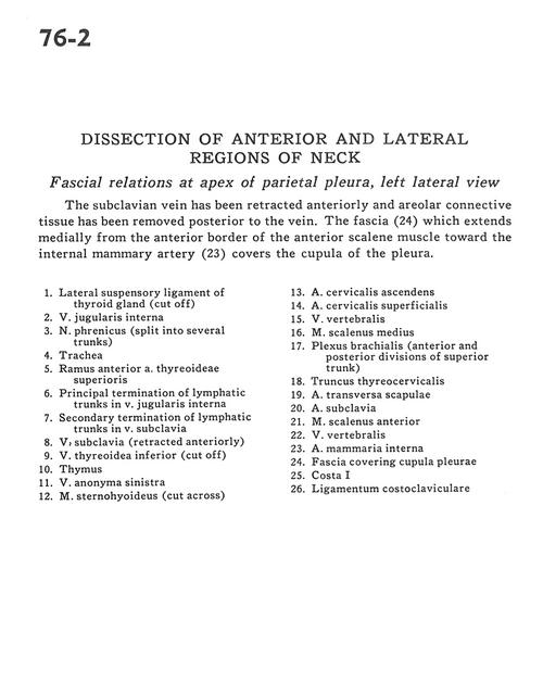
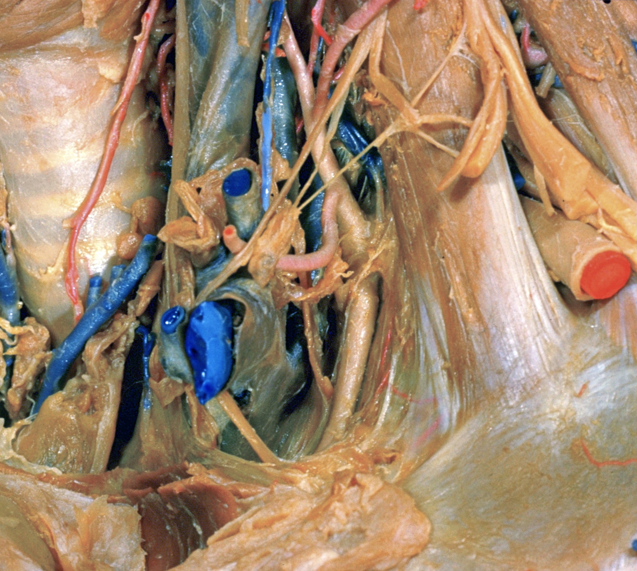
Dissection of anterior and lateral regions of neck
Fascial relations at apex of parietal pleura, left lateral view
The subclavian vein has been retracted anteriorly and areolar connective tissue has been removed posterior to the vein. The fascia (24) which extends medially from the anterior border of the anterior scalene muscle toward the internal mammary artery (23) covers the cupula of the pleura.
- Lateral suspensory ligament of thyroid gland (cut off)
- Internal jugular vein
- Phrenic nerve (split into several trunks)
- Trachea
- Anterior branch superior thyroid artery
- Principal termination of lymphatic trunks in internal jugular vein
- Secondary termination of lymphatic trunks in subclavian vein
- Subclavian vein (retracted anteriorly)
- Inferior thyroid vein (cut off)
- Thymus
- Brachiocephalic vein left
- Sternohyoid muscle (cut across)
- Ascending cervical artery
- Superficial cervical artery
- Vertebral vein
- Middle scalene muscle
- Brachial plexus (anterior and posterior divisions of superior trunk)
- Thyrocervical trunk
- Transverse scapular artery
- Subclavian artery
- Anterior scalene muscle
- Vertebral vein
- Internal thoracic (mammary) artery
- Fascia covering cupula pleurae
- Rib I
- Costoclavicular ligament