X CLOSE

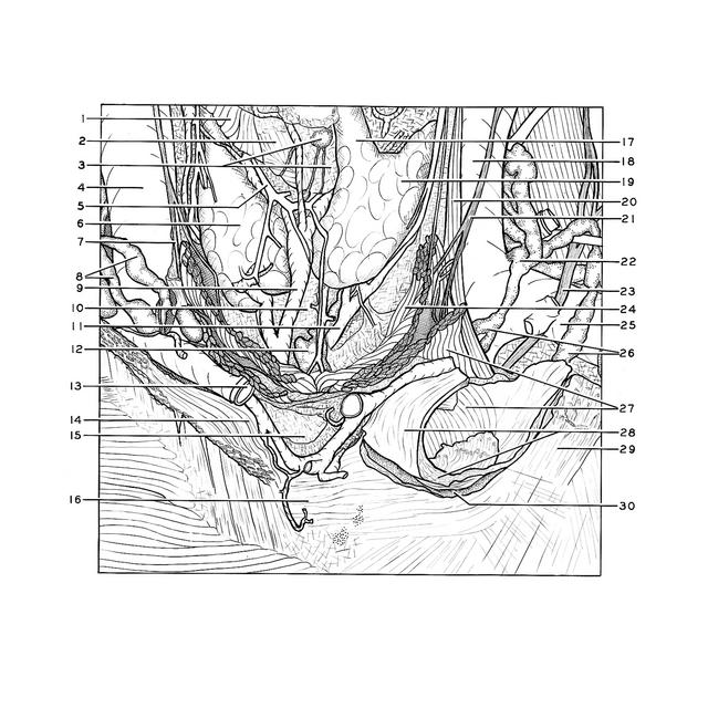
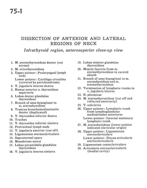
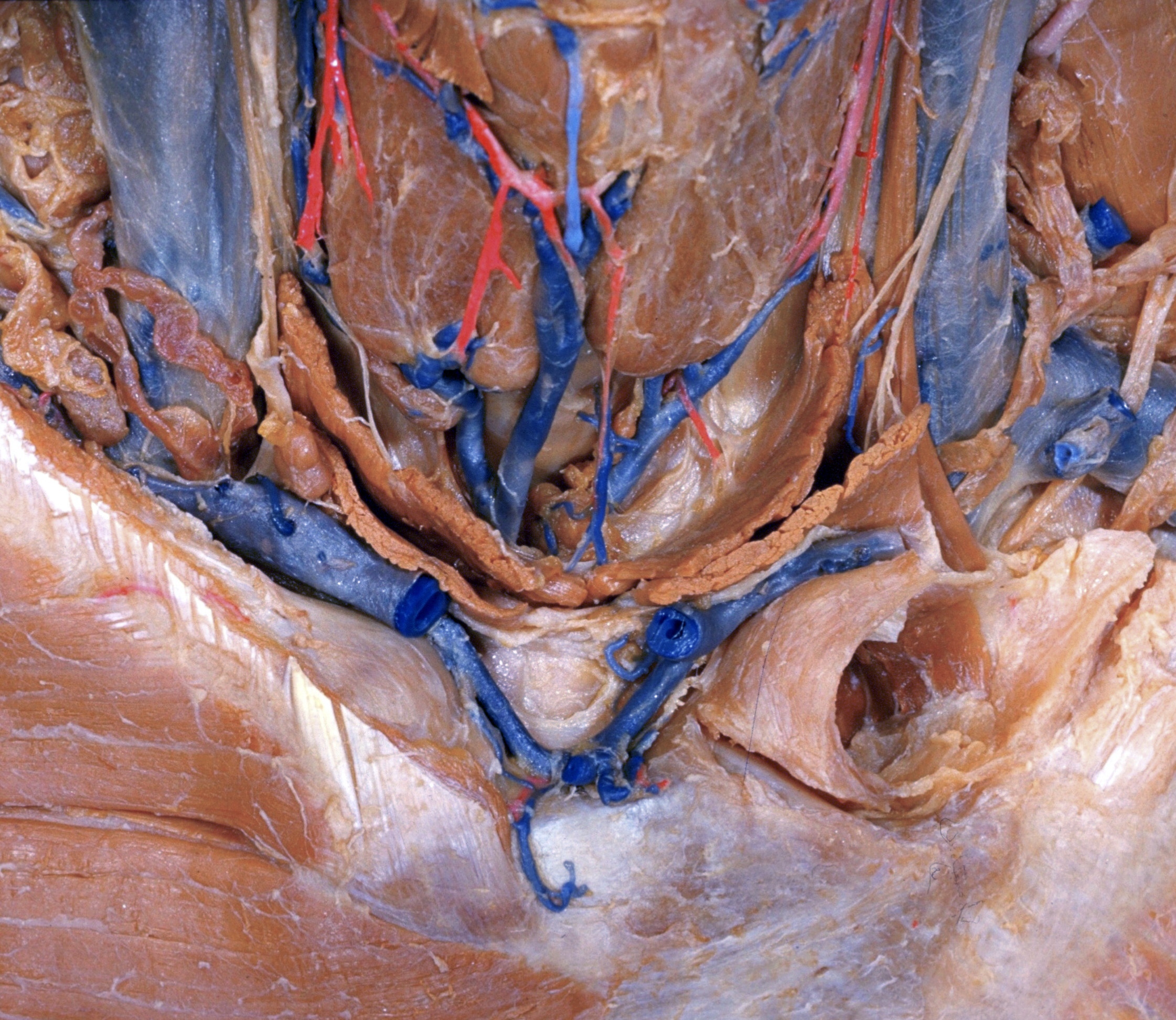
Dissection of anterior and lateral regions of neck

Infrahyoid region, anterosuperior close-up view

Stanford holds the copyright to the David L. Bassett anatomical images and has assigned Creative Commons license Attribution-Share Alike 4.0 International to all of the images.
For additional information regarding use and permissions, please contact Dr. Drew Bourn at dbourn@stanford.edu.

Image #75-1
Bassett ImageDiagram Image
Legend Image
Dissection of anterior and lateral regions of neck
Infrahyoid region, anterosuperior close-up view
1 . Sternothyroid muscle right (cut across)
2 . Cricothyroid muscle
3 . Upper pointer: Prelaryngeal lymph node Lower pointer: Cricoid cartilage (covered by perichondrium)
4 . Internal jugular vein right
5 . Branch anterior superior thyroid artery
6 . Right lobe of thyroid gland
7 . Branch of ansa hypoglossi to sternohyoid muscle
8 . Brachiocephalic trunk right (duplicated)
9 . Inferior thyroid vein right
10 . Trachea
11 . Inferior thyroid vein left
12 . Pretracheal lymph node
13 . Anterior jugular vein (cut off)
14 . Sternoclavicular ligament
15 . Suprasternal space
16 . Manubrium of sternum
17 . Pyramidal lobe of thyroid gland
18 . Internal jugular vein left
19 . Left lobe of thyroid gland
20 . Muscle fascicle from sternothyroid muscle to carotid sheath
21 . Branch of ansa hypoglossi to sternohyoid muscle and sternothyroid muscle
22 . Termination of lymphatic trunks in internal jugular vein
23 . Phrenic nerve
24 . Sternothyroid muscle (cut off and reflected anteriorly)
25 . Subclavian vein
26 . Upper pointer: Lymphatic trunk from anterior mediastinal lymph nodes Lower pointer: Internal mammary lymphatic trunk
27 . Sternohyoid muscle (lower pointer indicates clavicular origin)
28 . Upper pointer: Sternoclavicular ligament Lower pointer: Sternoclavicular articular disc
29 . Costoclavicular ligament
30 . Sternoclavicular joint (medial cavity)