Dissection of anterior and lateral regions of neck
Infrahyoid muscles, anterior view
Stanford holds the copyright to the David L. Bassett anatomical images and has assigned
Creative Commons license Attribution-Share
Alike 4.0 International to all of the images.
For additional information regarding use and permissions,
please contact Dr. Drew Bourn at dbourn@stanford.edu.
Image #73-6
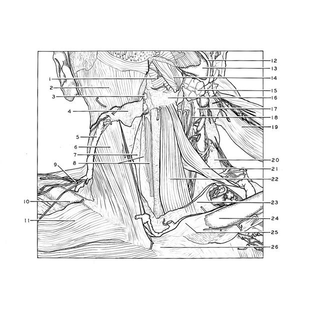
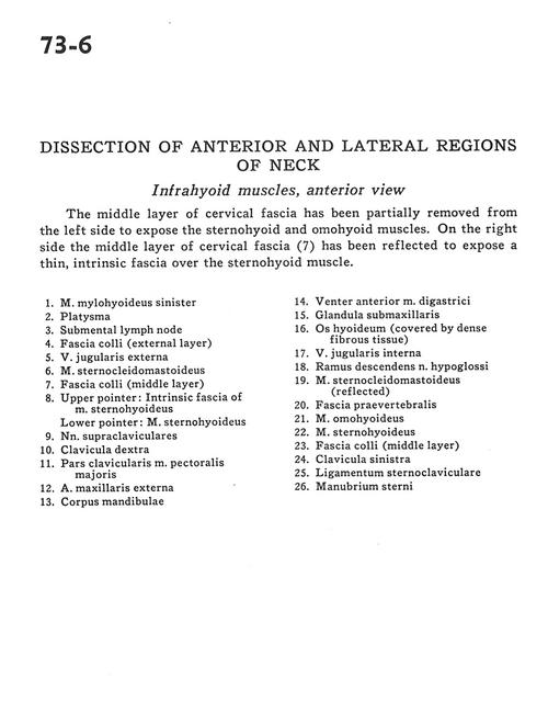
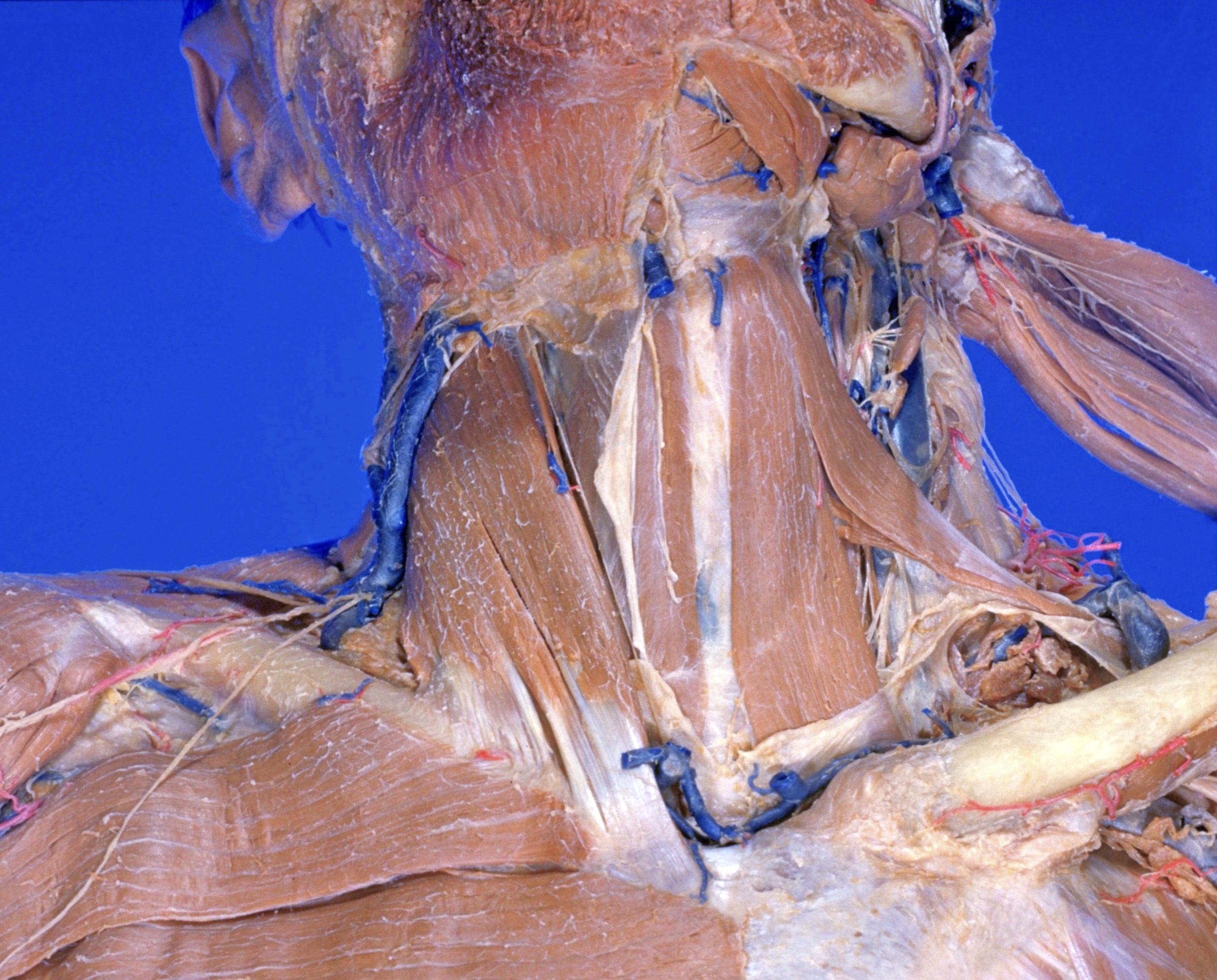
Dissection of anterior and lateral regions of neck
Infrahyoid muscles, anterior view
The middle layer of cervical fascia has been partially removed from the left side to expose the sternohyoid and omohyoid muscles. On the right side the middle layer of cervical fascia (7) has been reflected to expose a thin, intrinsic fascia over the sternohyoid muscle.
- Mylohyoid muscle left
- Platysma
- Submental lymph node
- Superficial fascia (external layer)
- External jugular vein
- Sternocleidomastoid muscle
- Superficial fascia (middle layer)
- Upper pointer: Intrinsic fascia of sternohyoid muscle Lower pointer: Sternohyoid muscle
- Supraclavicular nerves
- Clavicle right
- Clavicular part pectoralis major muscle
- External maxillary artery
- Body of mandible
- Anterior belly digastric muscle
- Submandibular gland
- Hyoid bone (covered by dense fibrous tissue)
- Internal jugular vein
- Descending branch hypoglossal nerve
- Sternocleidomastoid muscle (reflected)
- Prevertebral fascia
- Omohyoid muscle
- Sternohyoid muscle
- Superficial fascia (middle layer)
- Clavicle left
- Sternoclavicular ligament
- Manubrium of sternum