X CLOSE

Dissection of nasal fossae, nasal pharynx, and palate

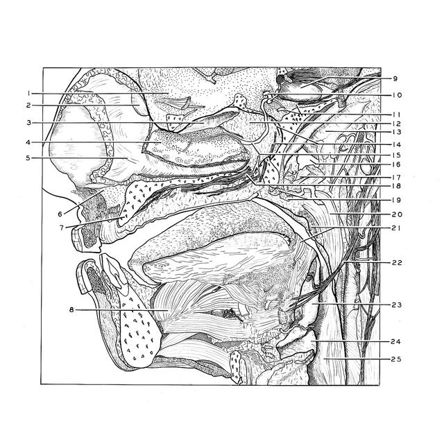
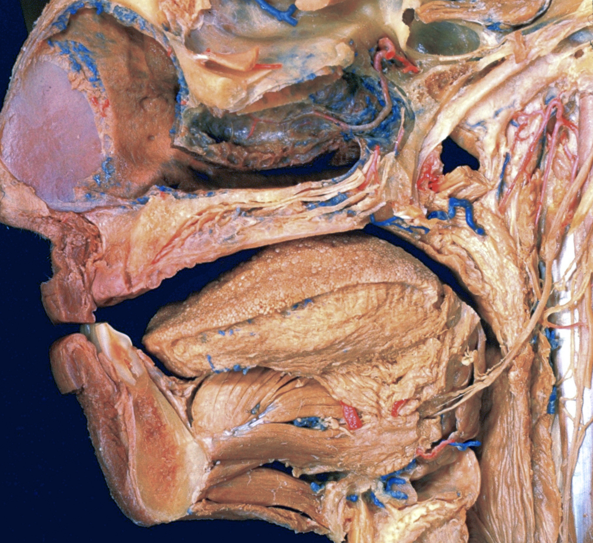
Blood vessels and nerves of lateral wall of left nasal fossa, lateral view

Stanford holds the copyright to the David L. Bassett anatomical images and has assigned Creative Commons license Attribution-Share Alike 4.0 International to all of the images.
For additional information regarding use and permissions, please contact Dr. Drew Bourn at dbourn@stanford.edu.

Image #71-6
Bassett ImageDiagram Image
Legend Image
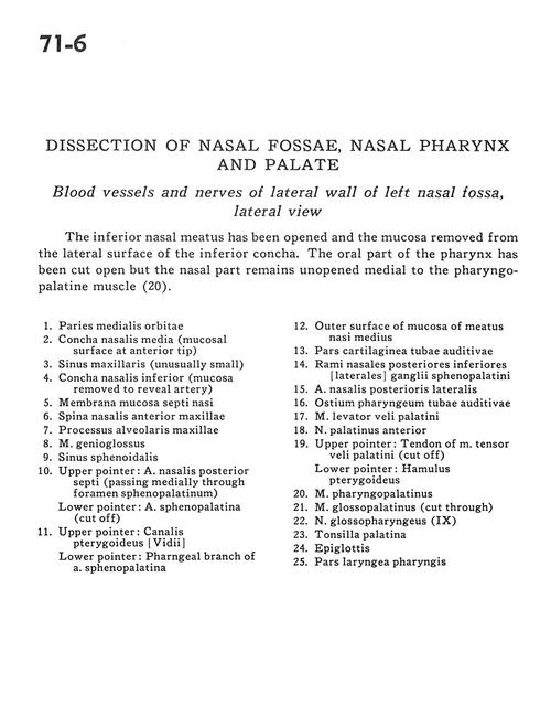
Dissection of nasal fossae, nasal pharynx, and palate
Blood vessels and nerves of lateral wall of left nasal fossa, lateral view
The inferior nasal meatus has been opened and the mucosa removed from the lateral surface of the inferior concha. The oral part of the pharynx has been cut open but the nasal part remains unopened medial to the pharyngopalatine muscle (20).
1 . Medial wall of orbit
2 . Middle nasal concha (mucosal surface at anterior tip)
3 . Maxillary sinus (unusually small)
4 . Inferior nasal concha (mucosa removed to reveal artery)
5 . Mucosal membrane nasal septum
6 . Anterior nasal spine maxilla
7 . Alveolar process of maxilla
8 . Genioglossus muscle
9 . Sphenoid sinus
10 . Upper pointer: Posterior nasal septal artery (passing medially through sphenopalatine foramen) Lower pointer: Sphenopalatine artery (cut off)
11 . Upper pointer: Pterygoid canal (Vidian) Lower pointer: Pharyngeal branch of sphenopalatine artery
12 . Outer surface of mucosa of middle nasal meatus
13 . Cartilaginous part auditory tube
14 . Posterior inferior nasal branches of sphenopalatine ganglion
15 . Posterior lateral nasal artery
16 . Bony pharyngeal auditory tube
17 . Levator veli palatini muscle
18 . Anterior palatine nerve
19 . Upper pointer: Tendon of tensor veli palatini muscle (cut off) Lower pointer: Pterygoid hamulus
20 . Pharyngopalatine muscle
21 . Glossopalatine muscle (cut through)
22 . Glossopharyngeal nerve (IX)
23 . Palatine tonsil
24 . Epiglottis
25 . Laryngeal part of pharynx