Exploration of the brain from its basal aspect
Relations of choroid plexus and fornix to transverse fissure
Stanford holds the copyright to the David L. Bassett anatomical images and has assigned
Creative Commons license Attribution-Share
Alike 4.0 International to all of the images.
For additional information regarding use and permissions,
please contact Dr. Drew Bourn at dbourn@stanford.edu.
Image #7-2
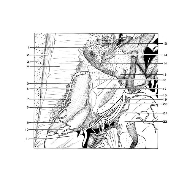
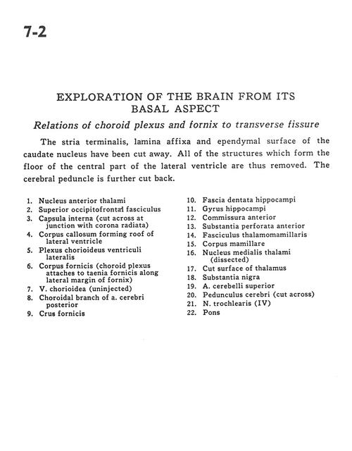
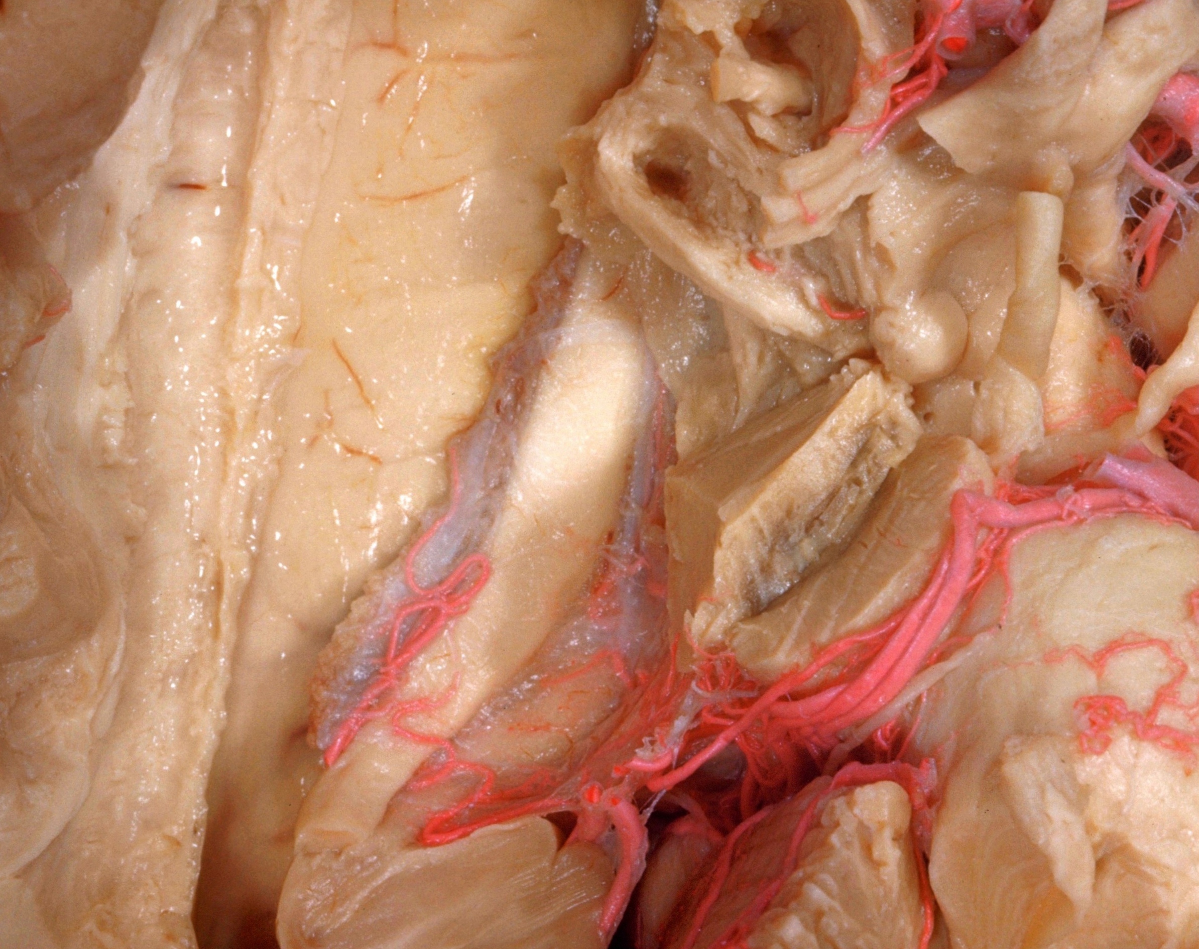
Exploration of the brain from its basal aspect
Relations of choroid plexus and fornix to transverse fissure
The stria terminalis, lamina affixa and ependymal surface of the caudate nucleus have been cut away. All of the structures which form the floor of the central part of the lateral ventricle are thus removed. The cerebral peduncle is further cut back.
- Anterior nucleus of thalamus
- Superior occipitofrontal fasciculus
- Internal capsule (cut across at junction with corona radiata)
- Corpus callosum forming roof of lateral ventricle
- Choroid plexus lateral ventricle
- Fornix (body) (choroid plexus attaches to head of fornix along lateral margin of fornix)
- Choroidal vein (uninjected)
- Choroidal branch of posterior cerebral artery
- Fornix (crus)
- Dentate fascia (hippocampus)
- Hippocampal gyrus
- Anterior commissure
- Anterior perforated substance
- Mamillothalamic tract
- Mamillary body
- Medial nucleus of thalamus (dissected)
- Cut surface of thalamus
- Substantia nigra
- Superior cerebellar artery
- Cerebral peduncle (cut across)
- Trochlear nerve (IV)
- Pons