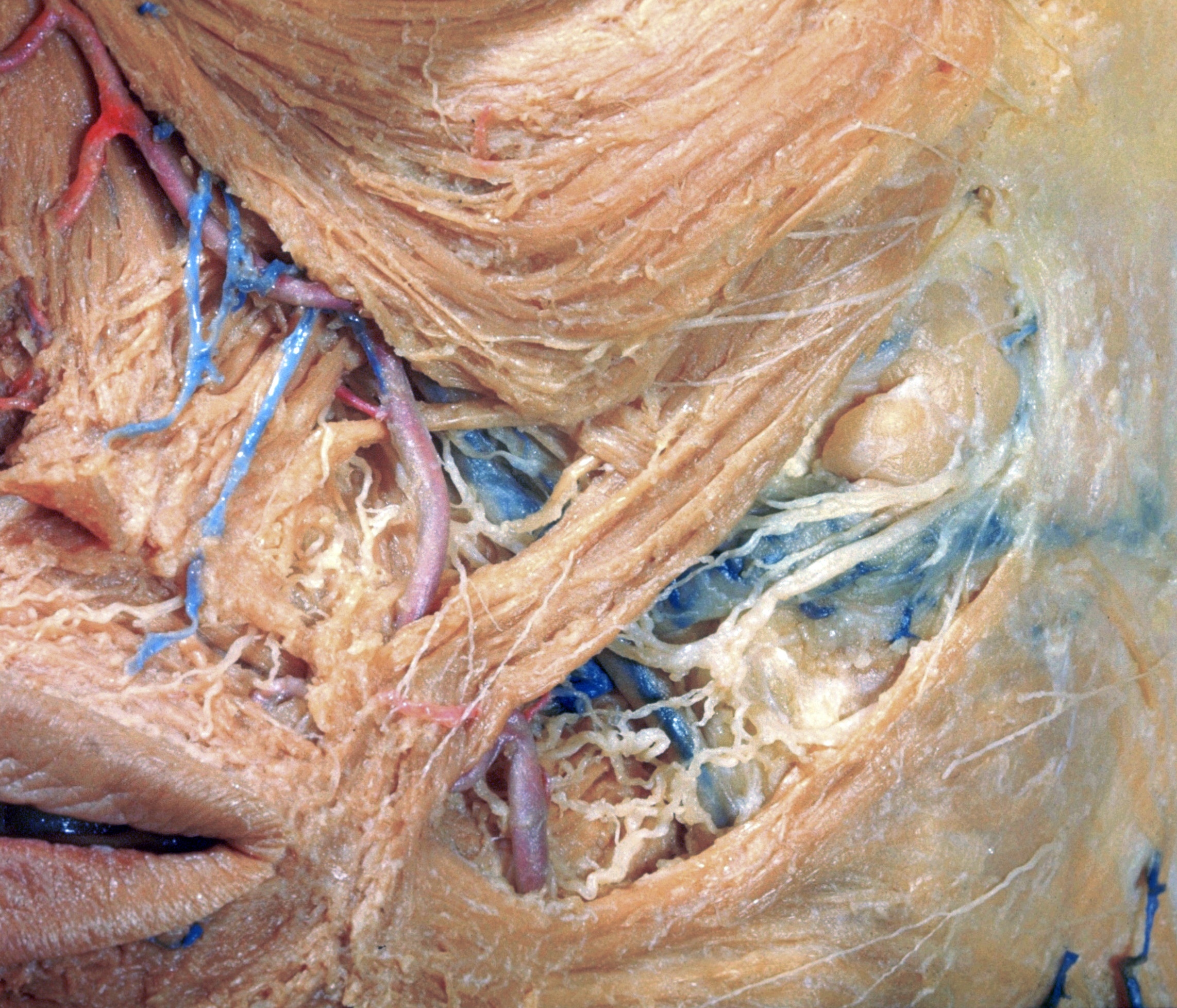
Left infraorbital, labial and buccal regions
Superficial nerves, blood vessels and muscles, anterolateral view
Stanford holds the copyright to the David L. Bassett anatomical images and has assigned
Creative Commons license Attribution-Share
Alike 4.0 International to all of the images.
For additional information regarding use and permissions,
please contact Dr. Drew Bourn at dbourn@stanford.edu.
Image #66-6
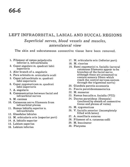
Left infraorbital, labial and buccal regions
Superficial nerves, blood vessels and muscles, anterolateral view
The skin and subcutaneous connective tissue have been removed.
- Filament of inferior palpebral branch of infraorbital nerve
- Angular head of levator labii superioris muscle
- Alar branch of angular artery
- Orbital part orbicularis oculi muscle
- Infraorbital head of levator labii superioris muscle
- Zygomatic head of levator labii superioris muscle
- Angular artery
- Communication between facial and infraorbital nerves
- Depressor anguli oris muscle
- Cutaneous nerve filaments from infraorbital plexus
- Superior labial branch of infraorbital nerve
- Labial glands
- Orbicularis oris muscle (superior part)
- Superior labial artery
- Upper lip
- Lower lip
- Orbicularis oris muscle (inferior part)
- Risorius muscle
- Zygomatic branches of facial nerve (several cutaneous filaments appear to be branches of the facial nerve although these are presumed to contain sensory fibers which reach the central nervous system through the trigeminal nerve)
- Accessory parotid gland
- Parotid-rnasseteric fascia
- Masseter muscle
- Buccal branch facial nerve (VII)
- Parotid duct (enclosed by sheath of connective tissue and plexus of veins)
- Zygomaticus muscle
- Anterior facial vein (incompletely filled with latex)
- External maxillary artery
- Filament of cutaneous colli nerve
- Buccinator muscle
- Platysma