Dissection of pharynx from left lateral approach
Auditory tube; levator veli palatini muscle, lateral view
Stanford holds the copyright to the David L. Bassett anatomical images and has assigned
Creative Commons license Attribution-Share
Alike 4.0 International to all of the images.
For additional information regarding use and permissions,
please contact Dr. Drew Bourn at dbourn@stanford.edu.
Image #66-4
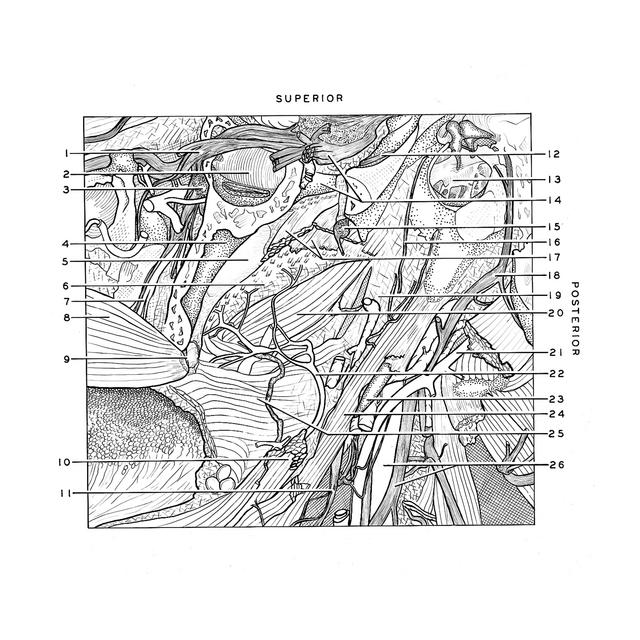
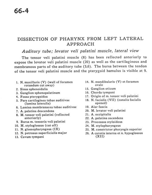
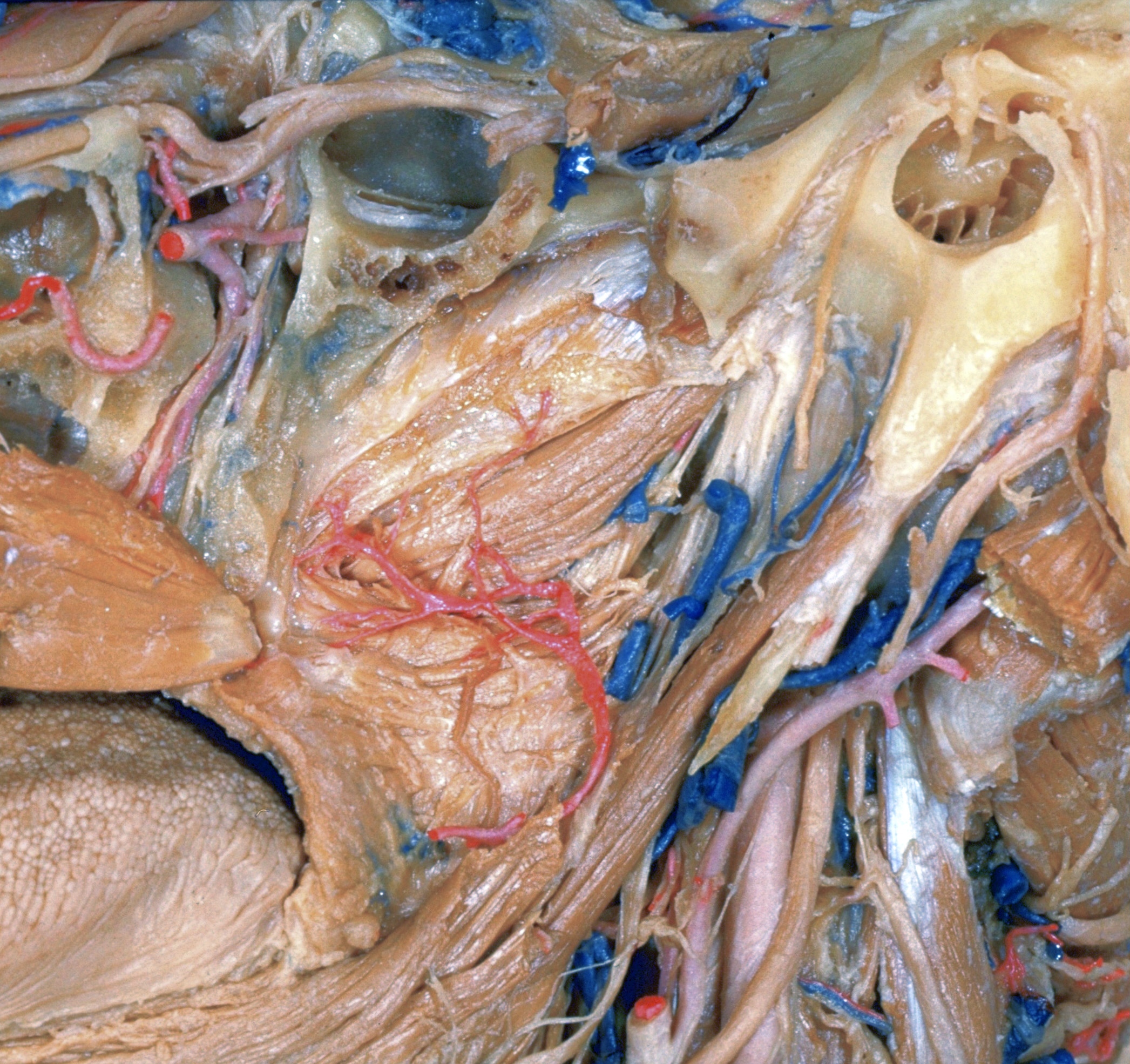
Dissection of pharynx from left lateral approach
Auditory tube; levator veli palatini muscle, lateral view
The tensor veli palatini muscle (8) has been reflected anteriorly to expose the levator veli palatini muscle (20) as well as the cartilaginous and membranous parts of the auditory tube(5,6). The bursa between the tendon of the tensor veli palatini muscle and the pterygoid hamulus is visible at 9.
- Maxillary nerve (V) (wall of foramen rotundum cut away)
- Sphenoid sinus
- Sphenopalatine ganglion
- Pterygoid fossa
- Cartilaginous part auditory tube (lateral plate)
- Membranous plate of auditory tube
- Descending palatine artery
- Tensor veli palatini muscle (reflected anteriorly)
- Bursa tensor veli palatini muscle
- Styloglossus muscle (cut off)
- Glossopharyngeal nerve (IX)
- Major superficial petrosal nerve
- Tympanic cavity
- Mandibular nerve (V) and foramen ovale
- Otic ganglion
- Chorda tympani
- Origin of tensor veli palatini muscle
- Facial nerve (VII) (facial canal opened)
- Alar fascia
- Levator veli palatini muscle
- Occipital artery
- Ascending palatine artery
- Styloid process
- Stylopharyngeus muscle
- Superior pharyngeal constrictor muscle
- Internal carotid artery and hypoglossal nerve (XII)