Dissection of left infratemporal and pterygopalatine fossae
Nerve supply to external pterygoid muscle; temporomandibular articulation, anterolateral view
Stanford holds the copyright to the David L. Bassett anatomical images and has assigned
Creative Commons license Attribution-Share
Alike 4.0 International to all of the images.
For additional information regarding use and permissions,
please contact Dr. Drew Bourn at dbourn@stanford.edu.
Image #65-4
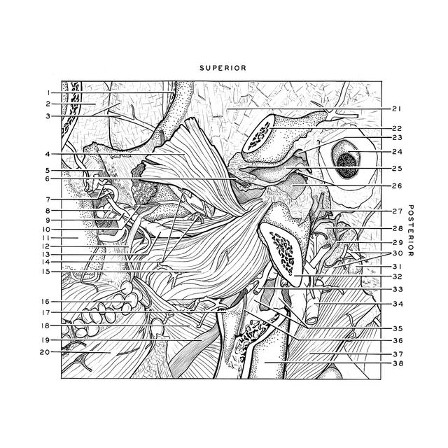
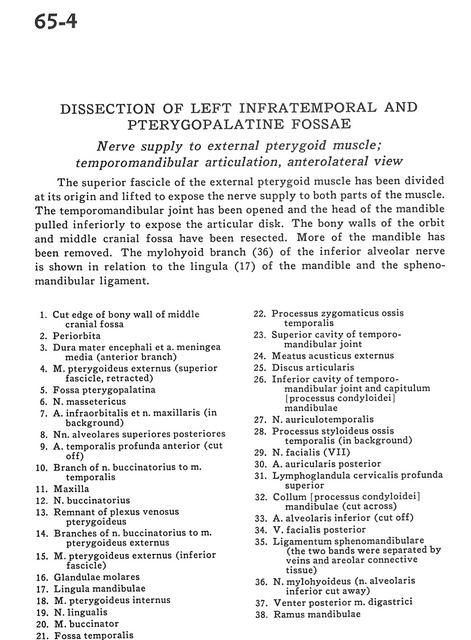
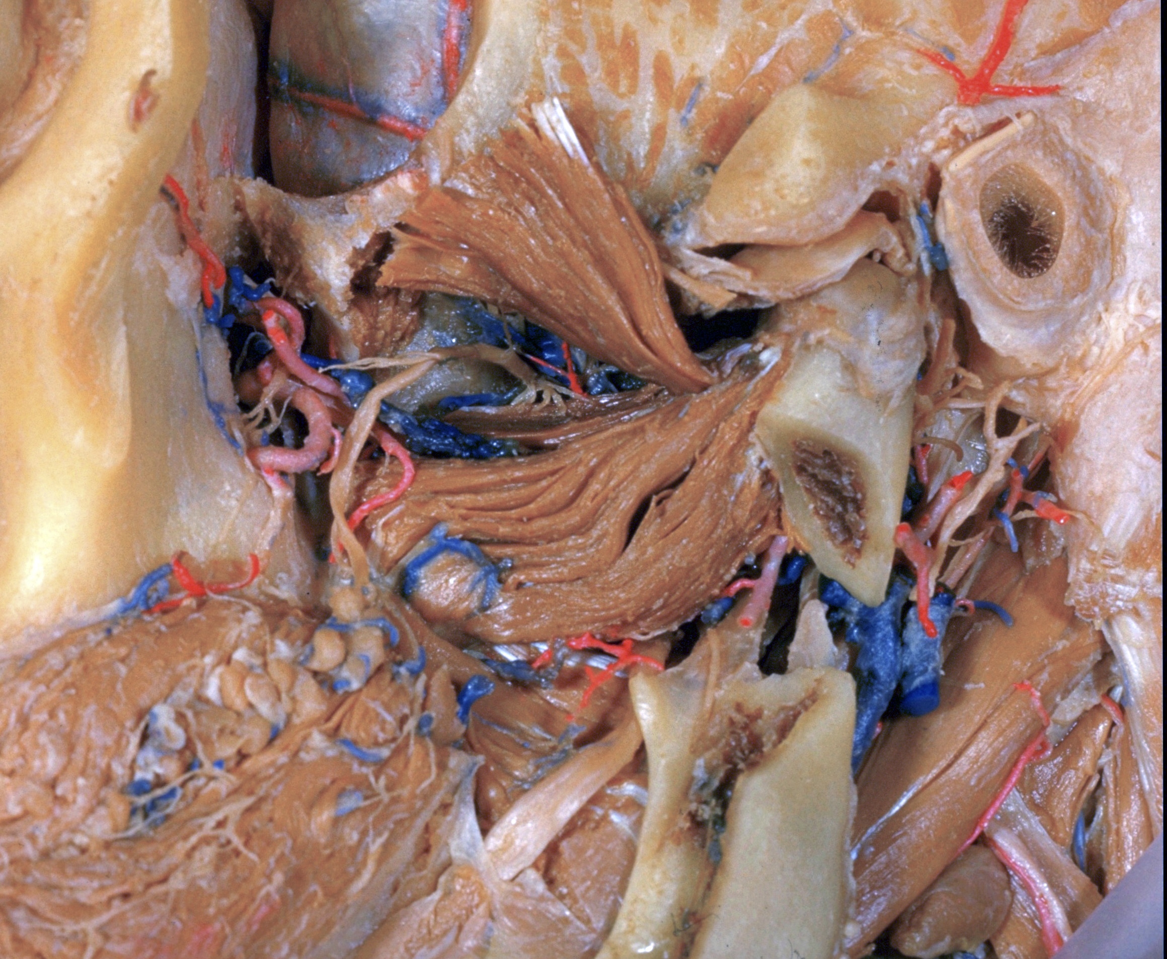
Dissection of left infratemporal and pterygopalatine fossae
Nerve supply to external pterygoid muscle; temporomandibular articulation, anterolateral view
The superior fascicle of the external pterygoid muscle has been divided at its origin and lifted to expose the nerve supply to both parts of the muscle. The temporomandibular joint has been opened and the head of the mandible pulled inferiorly to expose the articular disk. The bony walls of the orbit and middle cranial fossa have been resected. More of the mandible has been removed. The mylohyoid branch (36) of the inferior alveolar nerve is shown in relation to the lingula (17) of the mandible and the sphenomandibular ligament.
- Cut edge of bony wall of middle cranial fossa
- Periorbita
- Dura mater and middle meningeal artery (anterior branch)
- External pterygoid muscle (superior fascicle, retracted)
- Pterygopalatine fossa
- Masseteric nerve
- Infraorbital artery and maxillary nerve (in background)
- Superior posterior alveolar nerves
- Deep anterior temporal artery (cut off)
- Branch of buccinator nerve to temporalis muscle
- Maxilla
- Buccal nerve
- Remnant of pterygoid venous plexus
- Branches of buccinator nerve to external pterygoid muscle
- External pterygoid muscle (inferior fascicle)
- Molar glands
- Lingula of mandible
- Internal pterygoid muscle
- Lingual nerve
- Buccinator muscle
- Temporal fossa
- Zygomatic process temporal bone
- Superior cavity of temporomandibular joint
- Ext acoustic meatus
- Articular disc
- Inferior cavity of temporomandibular joint and capitulum (condyloid process) of mandible
- Auriculotemporal nerve
- Styloid process temporal bone (in background)
- Facial nerve (VII)
- Posterior auricular artery
- Deep cervical lymph node (superior)
- Neck (condyloid process) of mandible (cut across)
- Inferior alveolar artery (cut off)
- Posterior facial vein
- Sphenomandibular ligament (the two bands were separated by veins and areolar connective tissue)
- Mylohyoid nerve (inferior alveolar nerve cut away)
- Posterior belly of digastric muscle
- Ramus of mandible