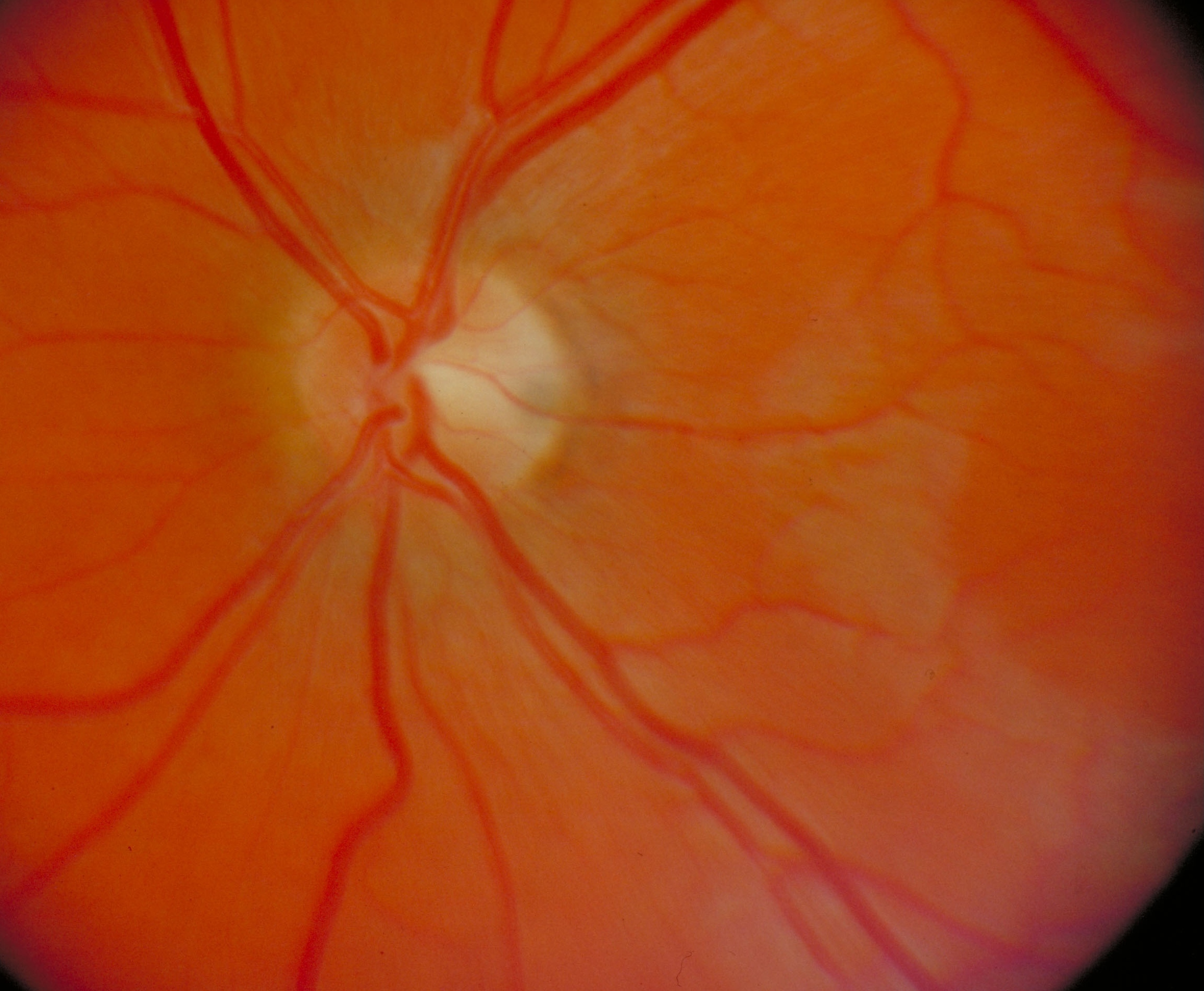
Fundus of left eye
Optic papilla, central retinary artery and vein, anterior view
Stanford holds the copyright to the David L. Bassett anatomical images and has assigned
Creative Commons license Attribution-Share
Alike 4.0 International to all of the images.
For additional information regarding use and permissions,
please contact Dr. Drew Bourn at dbourn@stanford.edu.
Image #59-1
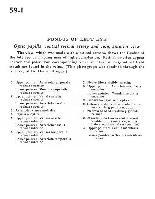
Fundus of left eye
Optic papilla, central retinary artery and vein, anterior view
The view, which was made with a retinal camera, shows the fundus of the left eye of a young man in light complexion. Retinal arteries appear narrow and paler than corresponding veins and have a longitudinal light streak not found in the veins. (This photograph was obtained through the courtesy of Dr. Homer Brugge.)
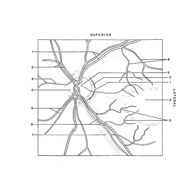
- Upper pointer: Superior temporal retinal artery Lower pointer: Superior temporal retinal vein
- Upper pointer: Superior nasal retinal vein Lower pointer: Superior retinal nasal artery
- Medial retinal artery
- Optic papilla
- Upper pointer: Inferior nasal retinal vein Lower pointer: Inferior nasal retinal artery
- Upper pointer: Inferior temporal retinal vein Lower pointer: Inferior temporal retinal artery
- Nerve fibers visible in retina
- Upper pointer: Superior macular artery Lower pointer: Superior macular vein
- Optic disc
- Sclera visible as narrow white zone surrounding optic papilla
- Narrow band of stratum pigmentosum
- Macula lutea (fovea centralis not visible in this instance; whitish halo around macula is common)
- Upper pointer: Inferior macular vein Lower pointer: Inferior macular artery