X CLOSE

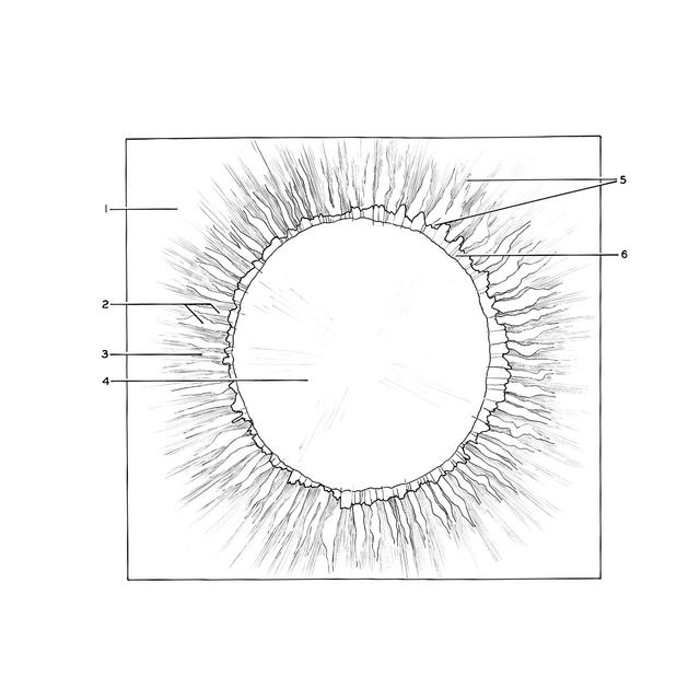
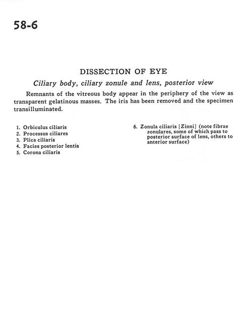
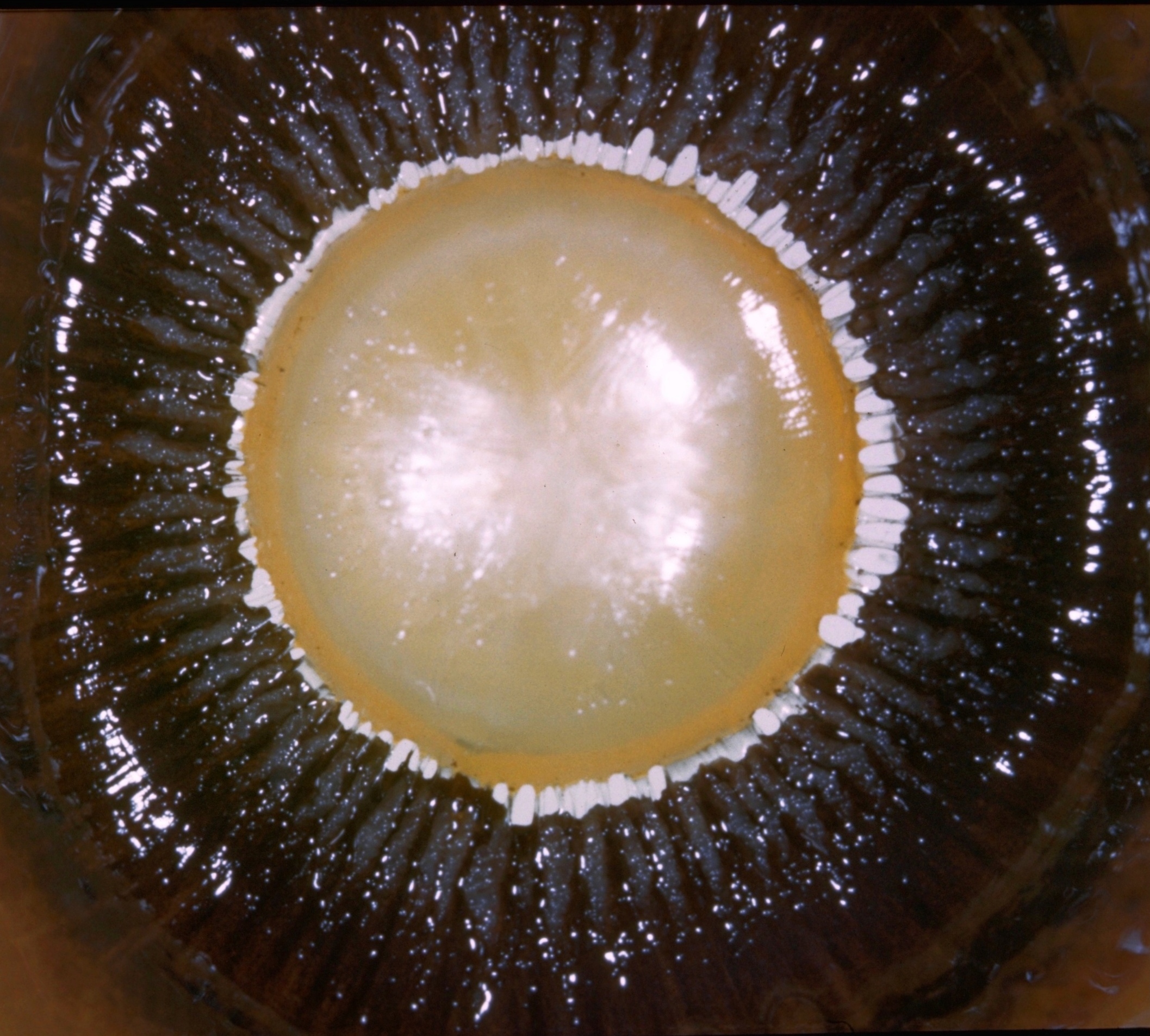
Dissection of eye

Ciliary body, ciliary zonule and lens, posterior view

Stanford holds the copyright to the David L. Bassett anatomical images and has assigned Creative Commons license Attribution-Share Alike 4.0 International to all of the images.
For additional information regarding use and permissions, please contact Dr. Drew Bourn at dbourn@stanford.edu.

Image #58-6
Bassett ImageDiagram Image
Legend Image
Dissection of eye
Ciliary body, ciliary zonule and lens, posterior view
Remnants of the vitreous body appear in the periphery of the view as transparent gelatinous masses. The iris has been removed and the specimen transilluminated.
1 . Orbiculus ciliaris
2 . Ciliary processes
3 . Plica ciliaris
4 . Posterior surface of lens
5 . Corona ciliaris
6 . Zonula ciliaris (note zonular fibers, some of which pass to posterior surface of lens, others to anterior surface)