Dissection of eye
Horizontal section of left eye
Stanford holds the copyright to the David L. Bassett anatomical images and has assigned
Creative Commons license Attribution-Share
Alike 4.0 International to all of the images.
For additional information regarding use and permissions,
please contact Dr. Drew Bourn at dbourn@stanford.edu.
Image #58-2
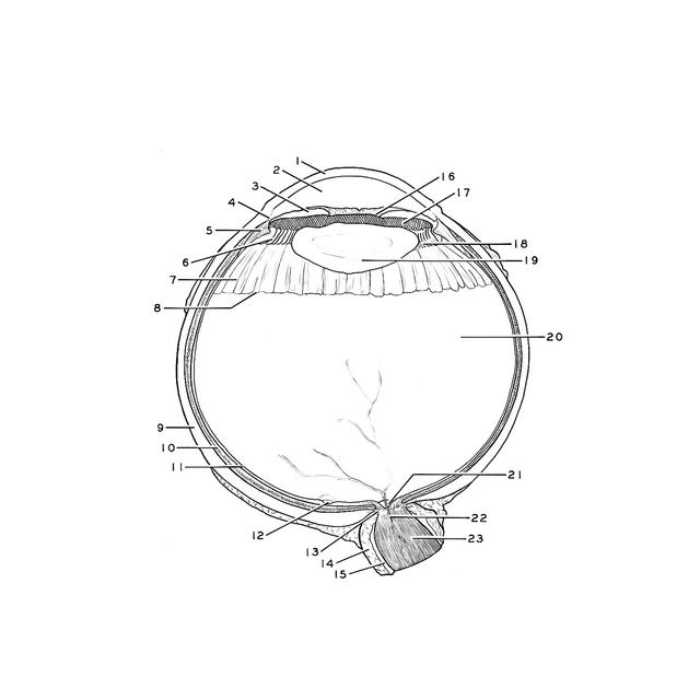
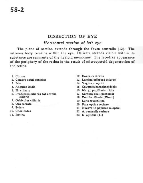
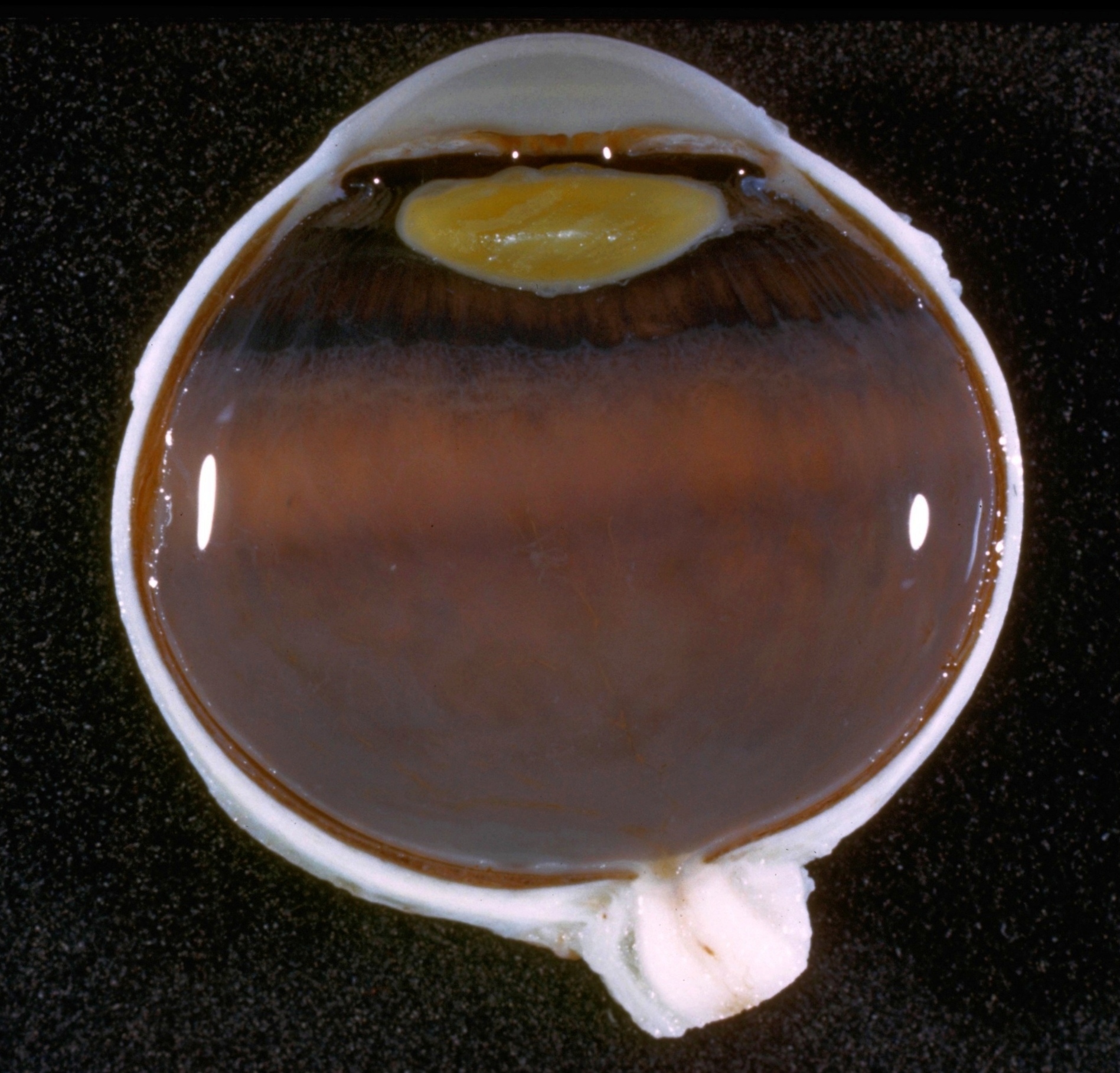
Dissection of eye
Horizontal section of left eye
The plane of section extends through fovea centralis(12). The vitreous body remains within the eye. Delicate strands visible within its substance are remnants of the hyaloid membrane. The lace-like appearance of the periphery of the retina is the result of microcystoid degeneration of the retina
- Cornea
- Anterior chamber of eye
- Iris
- Angle of iris
- Ciliary muscle
- Ciliary processes (of corona ciliaris)
- Orbiculus ciliaris
- Ora serrata
- Sclera
- Choroid
- Retina
- Fovea centralis
- Cribriform plate of sclera
- Sheath of optic nerve
- Subarachnoid space
- Iris-pupillary margin
- Posterior chamber of eye
- Zonula ciliaris [Zinn]
- Crystalline lens
- Optic (visual) part of retina
- Optic disc
- Central artery of retina
- Optic nerve (II)