X CLOSE

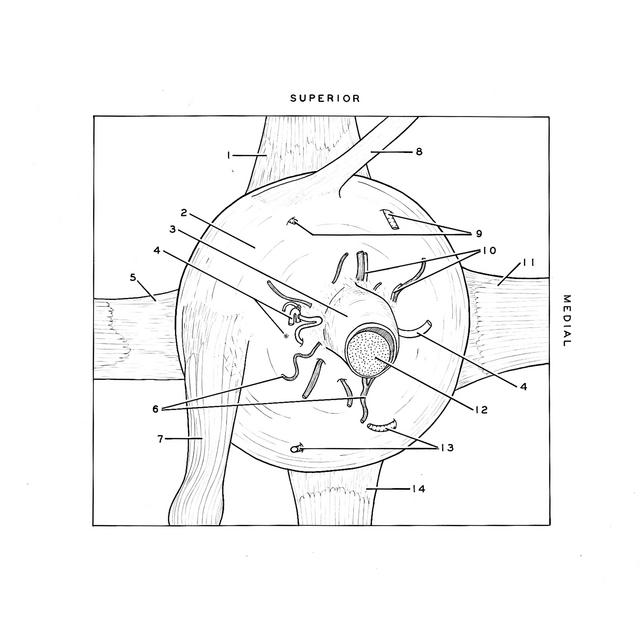
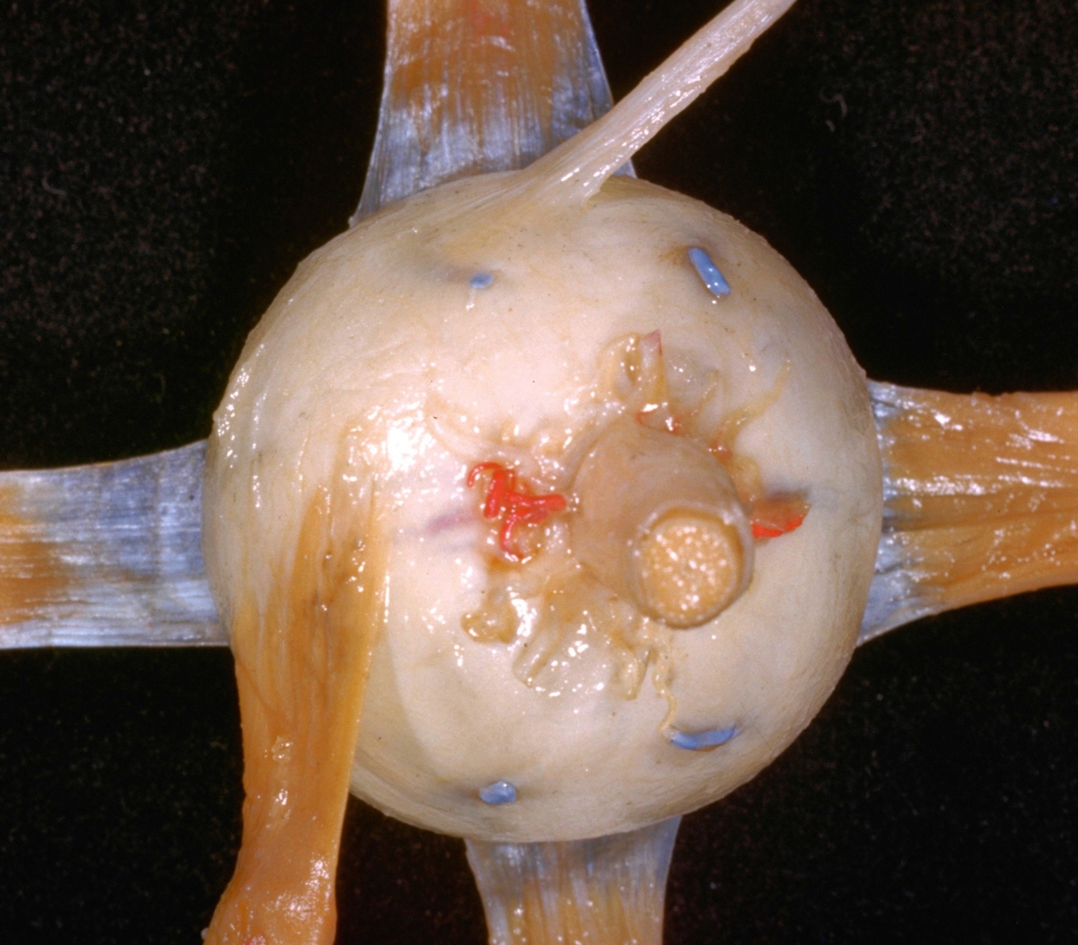
Insertion of extraocular muscles

Left eye, posterior view

Stanford holds the copyright to the David L. Bassett anatomical images and has assigned Creative Commons license Attribution-Share Alike 4.0 International to all of the images.
For additional information regarding use and permissions, please contact Dr. Drew Bourn at dbourn@stanford.edu.

Image #57-6
Bassett ImageDiagram Image
Legend Image
Insertion of extraocular muscles
Left eye, posterior view
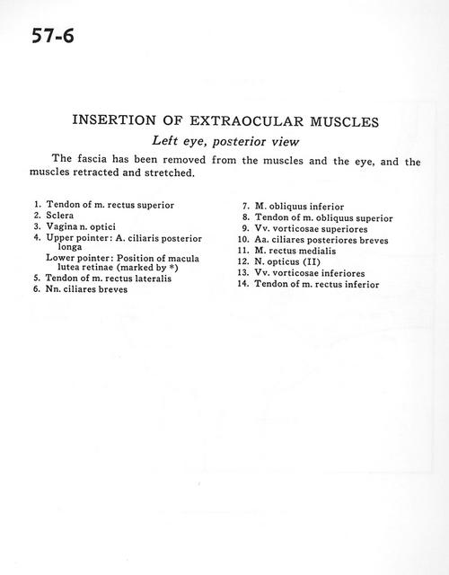
The fascia has been removed from the muscles and the eye, and the muscles retracted and stretched.
1 . Tendon of superior rectus muscle
2 . Sclera
3 . Sheath of optic nerve
4 . Upper pointer: Posterior ciliary artery (long) Lower pointer: Position of macula lutea of retina (marked by *)
5 . Tendon of lateral rectus muscle
6 . Short ciliary nerves
7 . Inferior oblique muscle
8 . Tendon of superior oblique muscle
9 . Superior vorticose veins
10 . Posterior ciliary arteries (short)
11 . Medial rectus muscle
12 . Optic nerve (II)
13 . Inferior vorticose veins
14 . Tendon of Inferior rectus muscle