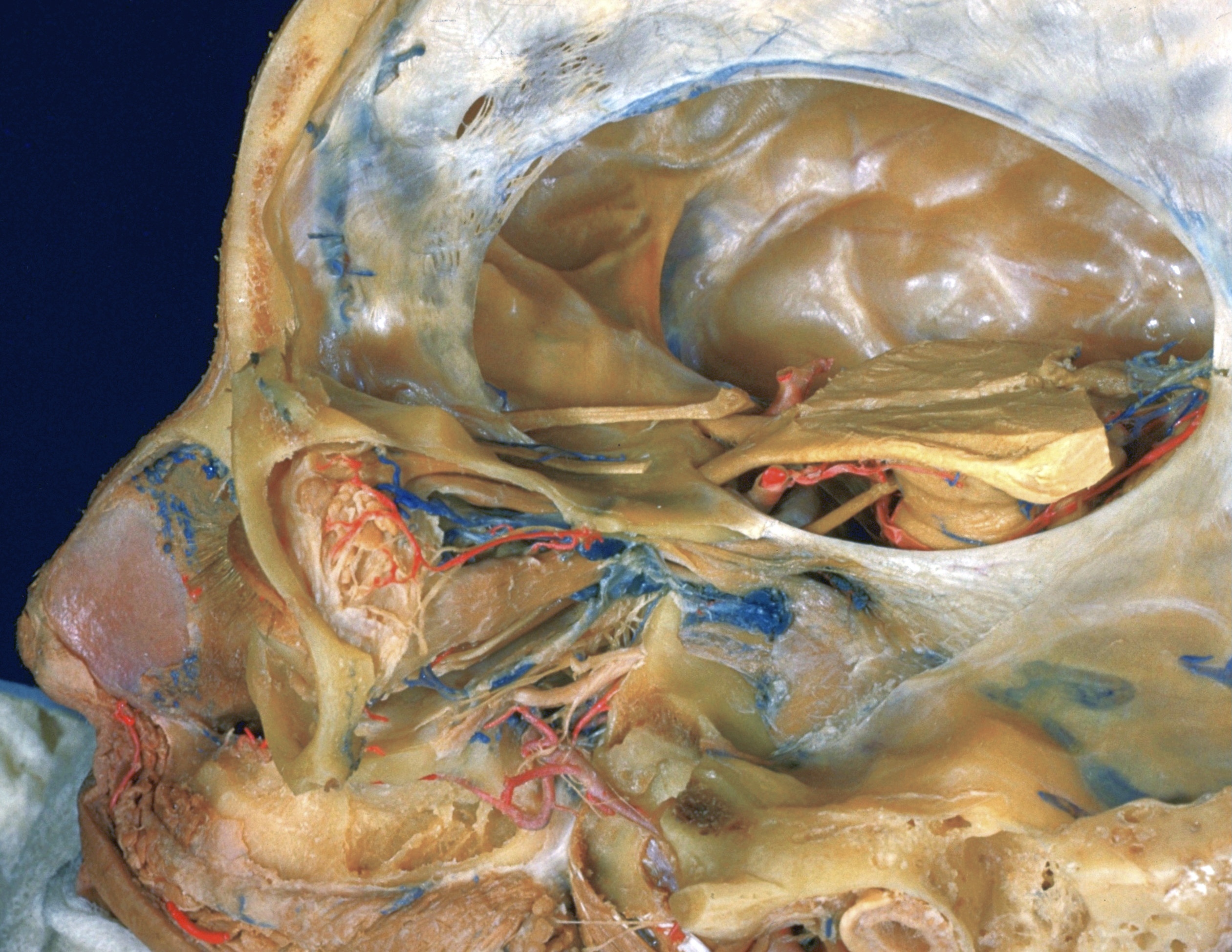
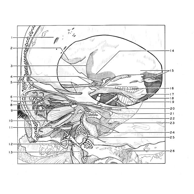
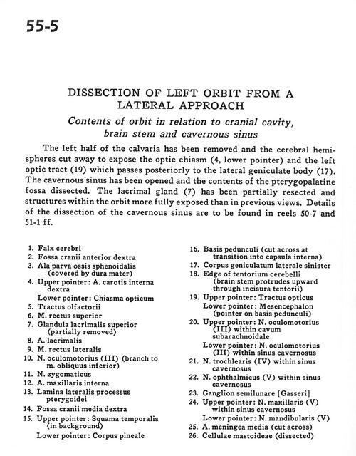
Dissection of left orbit from a lateral approach
Contents of orbit in relation to cranial cavity, brain stem and cavernous sinus
Stanford holds the copyright to the David L. Bassett anatomical images and has assigned
Creative Commons license Attribution-Share
Alike 4.0 International to all of the images.
For additional information regarding use and permissions,
please contact Dr. Drew Bourn at dbourn@stanford.edu.
Image #55-5
Dissection of left orbit from a lateral approach
Contents of orbit in relation to cranial cavity, brain stem and cavernous sinus
The left half of the calvaria has been removed and the cerebral hemispheres cut away to expose the optic chiasm (4, lower pointer) and the left optic tract(19) which passes posteriorly to the lateral geniculate body (17). The cavernous sinus has been opened and the contents of the pterygopalatine fossa dissected. The lacrimal gland (7) has been partially resected and structures within the orbit more fully exposed than in previous views. Details of the dissection of the cavernous sinus are to be found in reels 50-7 and 51-1 ff.
- Falx cerebri
- Anterior cranial fossa right
- Lesser wing sphenoid bone (covered by dura mater)
- Upper pointer: Internal carotid artery right Lower pointer: Optic chiasm
- Olfactory tracts
- Superior rectus muscle
- Superior lacrimal gland (partially removed)
- Lacrimal artery
- Lateral rectus muscle
- Oculomotor nerve (III) (branch to inferior oblique muscle)
- Zygomatic nerve
- Internal maxillary artery
- Lateral plate of pterygoid process
- Middle cranial fossa right
- Upper pointer: Temporal bone (squamous part) (in background) Lower pointer: Pineal body
- Base of peduncle (cut across at transition into internal capsule)
- Lateral geniculate body left
- Edge of tentorium cerebelli (brain stem protrudes upward through tentorial incisure)
- Upper pointer: Optic tract Lowerpointer: Mesencephalon (pointer on base of peduncle)
- Upper pointer: Oculomotor nerve (III) within subarachnoid space Lower pointer: Oculomotor nerve (II) within cavernous sinus
- Trochlear nerve (IV) within cavernous sinus
- Ophthalmic nerve (V) within cavernous sinus
- Semilunar ganglion (trigeminal)
- Upper pointer: Maxillary nerve (V) within cavernous sinus Lower pointer: Mandibular nerve (V)
- Middle meningeal artery (cut across)
- Mastoid cells (dissected)