Scalp
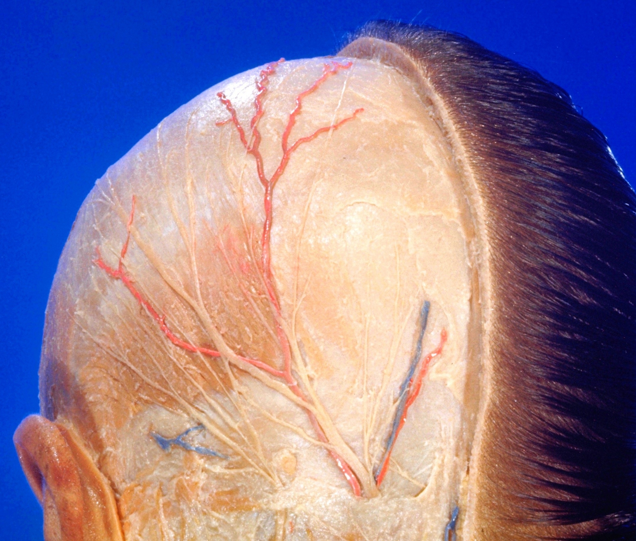
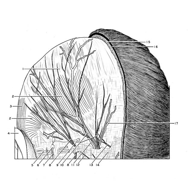
Superficial nerves and blood vessels of scalp, posterior view
Stanford holds the copyright to the David L. Bassett anatomical images and has assigned
Creative Commons license Attribution-Share
Alike 4.0 International to all of the images.
For additional information regarding use and permissions,
please contact Dr. Drew Bourn at dbourn@stanford.edu.
Image #51-7
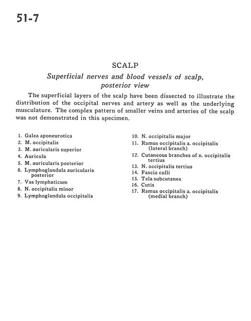
Scalp
Superficial nerves and blood vessels of scalp, posterior view
The superficial layers of the scalp have been dissected to illustrate the distribution of the occipital nerves and artery as well as the underlying musculature. The complex pattern of smaller veins and arteries of the scalp was not demonstrated in this specimen.
- Galea aponeurotica
- Occipitalis muscle
- Superior auricular muscle
- Auricle
- Posterior auricular muscle
- Posterior auricular lymph nodes
- Lymph vessel
- Lesser occipital nerve
- Occipital lymph nodes
- Occipital major nerve
- Occipital branch of occipital artery (lateral branch)
- Cutaneous branches of third occipital nerve
- Third occipital nerve
- Superficial fascia
- Superficial fascia
- Scalp
- Occipital branch of occipital artery (medial branch)