X CLOSE

Osteology

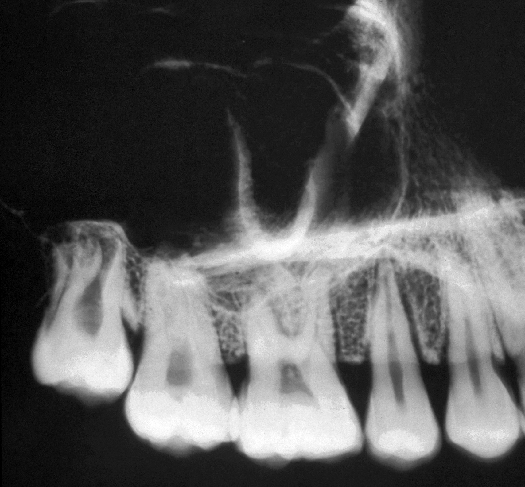
Right upper molar and premolar teeth, lateromedial roentgenogram

Stanford holds the copyright to the David L. Bassett anatomical images and has assigned Creative Commons license Attribution-Share Alike 4.0 International to all of the images.
For additional information regarding use and permissions, please contact Dr. Drew Bourn at dbourn@stanford.edu.

Image #47-4
Bassett ImageDiagram Image
Legend Image
Osteology
Right upper molar and premolar teeth, lateromedial roentgenogram
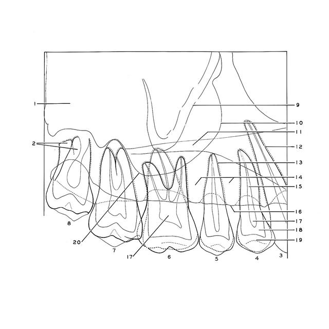
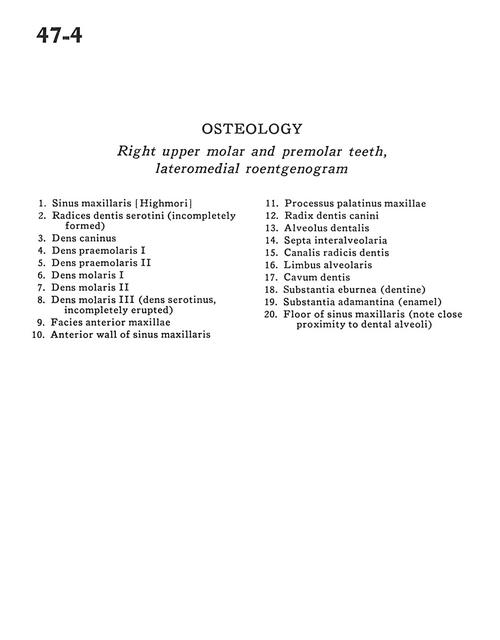
1 . Maxillary sinus (Highmore)
2 . Roots molar III (serotini) (incompletely formed)
3 . Canine
4 . Premolar I
5 . Premolar II
6 . Molar I
7 . Molar II
8 . Molar III (serotinus incompletely erupted)
9 . Anterior surface of maxilla
10 . Anterior wall of maxillary sinus
11 . Palatine process of maxilla
12 . Root of canine
13 . Alveolus
14 . Interalveolar septa
15 . Root canal
16 . Alveolar border
17 . Cavity
18 . Substantia ebumea (dentine)
19 . Substantia adamantina (enamel)
20 . Floor of maxillary sinus (note close proximity to dental alveoli)