X CLOSE

Osteology

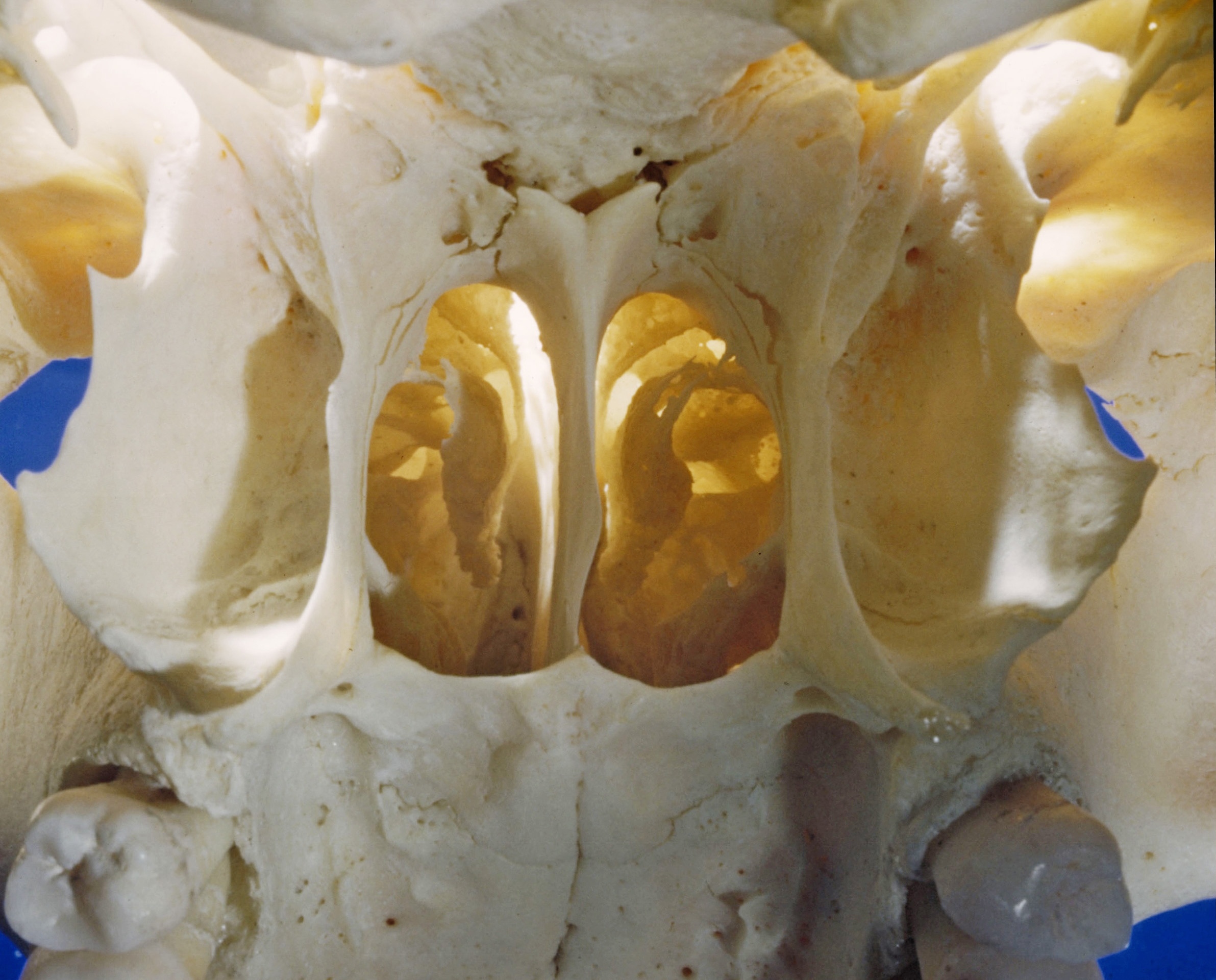
Nasal fossae, posterior view through choanae

Stanford holds the copyright to the David L. Bassett anatomical images and has assigned Creative Commons license Attribution-Share Alike 4.0 International to all of the images.
For additional information regarding use and permissions, please contact Dr. Drew Bourn at dbourn@stanford.edu.

Image #37-6
Bassett ImageDiagram Image
Legend Image
Osteology
Nasal fossae, posterior view through choanae
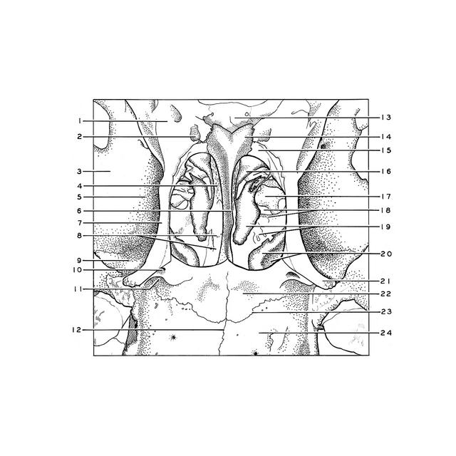
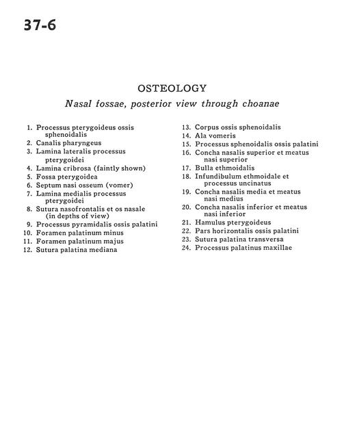
1 . Pterygoid process sphenoid bone
2 . Pharyngeal canal
3 . Lateral plate of pterygoid process
4 . Cribriform plate (faintly shown)
5 . Pterygoid fossa
6 . Bony nasal septum (vomer)
7 . Medial plate of pterygoid process
8 . Nasofrontal suture & nasal bone (in depths of view)
9 . Pyramidal process palatine bone
10 . Lesser palatine foramen
11 . Greater palatine foramen
12 . Median palatine suture
13 . Body sphenoid bone
14 . Ala of vomer
15 . Sphenoid process palatine bone
16 . Superior nasal concha & superior nasal meatus
17 . Ethmoidal bulla
18 . Infundibulum of ethmoid bone & uncinate process
19 . Middle nasal concha & middle nasal meatus
20 . Inferior nasal concha &Inferior nasal meatus
21 . Pterygoid hamulus
22 . Horizontal plate palatine bone
23 . Transverse palatine suture
24 . Palatine process of maxilla