Osteology
Roentgenogram of left orbit and optic canal
Stanford holds the copyright to the David L. Bassett anatomical images and has assigned
Creative Commons license Attribution-Share
Alike 4.0 International to all of the images.
For additional information regarding use and permissions,
please contact Dr. Drew Bourn at dbourn@stanford.edu.
Image #37-2
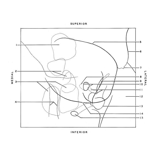
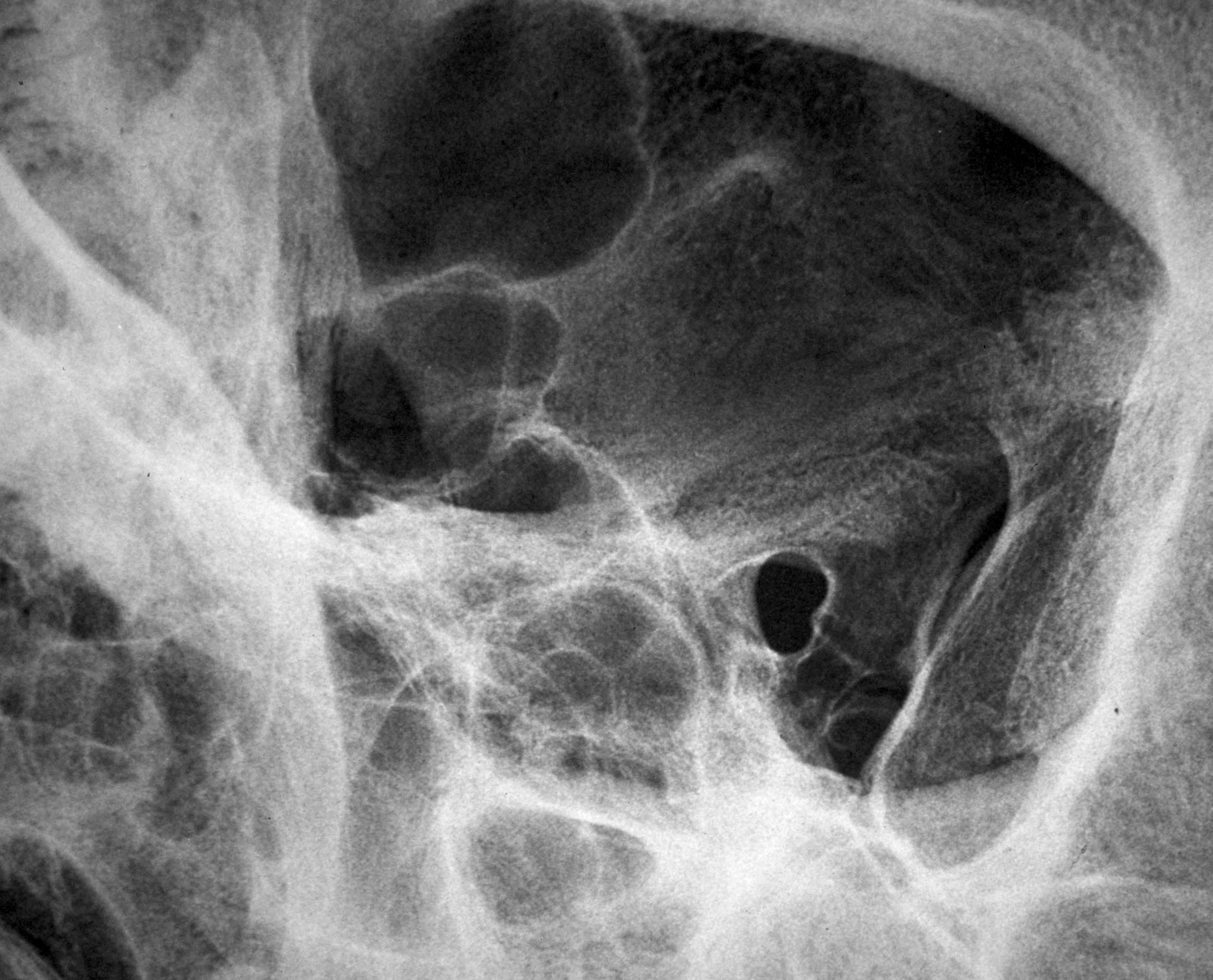
Osteology
Roentgenogram of left orbit and optic canal
This is an anterior view with an upward and medial angulation in line with the axis of the optic canal. The posterior portion of the skull has been cut away to avoid conflicting shadows. The relation of the anterior clinoid process (11) to the optic canal (10) is shown.
- Frontal sinus (ethmofrontal)
- Ethmoidal cells (along medial wall of orbit)
- Sphenoidal sinus (in bâckground)
- Borders of Nasolacrimal canal (in foreground)
- Supraorbital margin
- Temporal line of frontal bone
- Zygomaticofrontal suture
- Outline of posterior opening of optic canal
- Superior orbital fissure
- Outline of optic foramen (anterior opening of optic canal)
- Anterior clinoid process (lower pointer indicates tip of this process)
- Zygomatic bone
- Anterior limit of inferior orbital fissure
- Infraorbital margin
- Infraorbital foramen (maxillary sinus underlies floor of orbit in this region)