Osteology
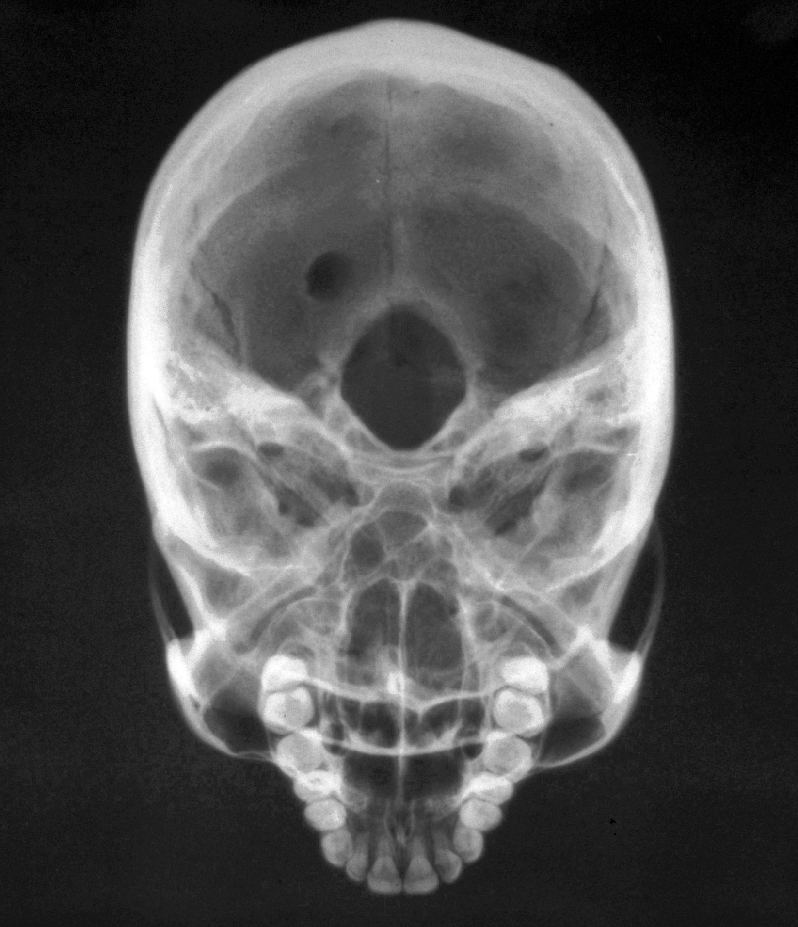
Roentgenogram of skull, inferosuperior view
Stanford holds the copyright to the David L. Bassett anatomical images and has assigned
Creative Commons license Attribution-Share
Alike 4.0 International to all of the images.
For additional information regarding use and permissions,
please contact Dr. Drew Bourn at dbourn@stanford.edu.
Image #35-7

Osteology
Roentgenogram of skull, inferosuperior view
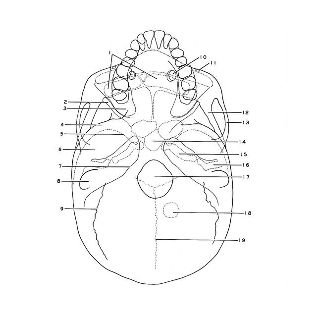
In this specimen one of the depressions (18) in the parietal bone which accommodates arachnoidal granulations (see Section I, 1-3) is unusually prominent as an area of rarification in the bone.
- Frontal sinus
- Inferior orbital fissure
- Maxillary sinus (posterior portion)
- Wall of cranial vault separating middle cranial fossa (below) from infratemporal fossa (above)
- Lesser wing of sphenoid bone (note continuation medially into anterior clinoid process)
- Position of mandibular fossa
- Posterior margin of petrosal part of temporal bone
- Mastoid process
- Occipitomastoid suture
- Nasolacrimal canal
- Anterior surface of maxilla (pointer near Infraorbital foramen)
- Lateral wall of cranial vault (anterior cranial fossa lies medial to this)
- Zygomatic arch
- Sphenoid sinus (ethmoidal cells visible anteriorly)
- Carotid canal
- External acoustic meatus
- Foramen magnum occipital (junction of coronal suture and sagittal suture visible in background)
- Depression in parietal bone for arachnoidal granulations
- Sagittal suture